
Object Detection as Campylobacter Bacteria and Phagocytotic Activity
of Leukocytes in Gram Stained Smears Images
Kyohei Yoshihara and Kouich Hirata
Kyushu Institute of Technology, Kawazu 680-4, Iizuka 820-8502, Japan
Keywords:
Object Detection, Campylobacter Bacteria, Phagocytotic Activity of Leukocytes, Gram Stained Smears
Images, Faster R-CNN, RetinaNet, YOLOv5.
Abstract:
In this paper, we apply object detection to Gram stained smear images, where objects are Campylobacter
bacteria and phagocytotic activity of leukocytes. Then, we adopt three CNN-based object detectors of Faster
R-CNN, RetinaNet and YOLOv5. The outline of the detection is first to annotate the regions of objects as
Campylobacter bacteria and phagocytotic activity of leukocytes in training images, and then to detect the
regions of objects in the remained test images by using the detectors. Finally, we give experimental results of
detecting Campylobacter bacteria and phagocytotic activity of leukocytes in Gram stained smear images by
using the detectors.
1 INTRODUCTION
The Gram stain (Bartholomew and Mittwer, 1952) is
the method for microbial smears test in microscope
test per 1, 000× field, introduced by Hans Christian
Gram (1853–1938)at 1884. For the Gram stain, based
on the stained colors as purple/violet or red/pink, the
stained shapes as sphere-shape, rod-shape, singles,
pairs, chains, clusters, and so on, we detect bacteria
occurring in the smears for the samples of blood, spu-
tum, feces, pus and urine.
After Gram staining, we call the bacteria colored
by purple or violet Gram positive and those by red or
pink Gram negative. Also we call the bacteria stained
as sphere-shape cocci and those as rod-shape basilli.
Hence, we can classify bacteria into the four kinds
as Gram positive cocci, Gram positive bacilli, Gram
negative cocci and Gram negative bacilli
1
.
Since the Gram stain is applicable inexpensively
and fast returns the results (within 30 min.), it is im-
portant for the initial medical care of infectious dis-
eases. On the other hand, Gram staining is possible
to stain not only bacteria but also non-bacteria sub-
stances such as leukocytes, dusts, oil and crystals.
Also there exist many kinds of phlogogenic bacteria
for infectious diseases.
In the microscope test, Gram stained smears im-
ages are checked manually and visually but not auto-
1
Sometimes we call basilli rods (Smith et al., 2018).
matically in general. The reason is that we can detect
bacteria exactly by applying the culture test and the
identification test after the microscope test. On the
other hand, since anaerobic bacteria are never lived in
the culture test, they cannot be detected by the iden-
tification test. Then, the detected bacteria through the
culture test and the identification test are the part of
bacteria in smears.
Also since the culture test and the identification
test spend one day, we cannot apply them to the ini-
tial medical care of infectious diseases. Furthermore,
whereas expert skills are necessary to detect bacte-
ria manually and visually from Gram stained smears
images, such technicians with expert skills are not
enough to apply the initial medical care in Japan.
Hence, the automatic detection of bacteria from Gram
stained smears images is required.
In this paper, we focus on Campylobacterbacteria
and phagocytotic activity of leukocytes. The Campy-
lobacter bacteria are Gram negative basilli and phlo-
gogenic bacteria causing so called Campylobacter en-
teritis. Also the phagocytotic activity of leukocytes
works as the natural immunity. Then, the detection of
them is an important tasks for the microscope test of
the Gram stained smears images. However, it is also
well-known to be difficult in the microscope test.
The difficulty of the detection of Campylobac-
ter bacteria is that we cannot distinguish the Campy-
lobacter bacteria from dusts, since the Campylobacter
bacteria are as small as dusts and their shapes are also
534
Yoshihara, K. and Hirata, K.
Object Detection as Campylobacter Bacteria and Phagocytotic Activity of Leukocytes in Gram Stained Smears Images.
DOI: 10.5220/0010813800003122
In Proceedings of the 11th International Conference on Pattern Recognition Applications and Methods (ICPRAM 2022), pages 534-541
ISBN: 978-989-758-549-4; ISSN: 2184-4313
Copyright
c
2022 by SCITEPRESS – Science and Technology Publications, Lda. All rights reserved
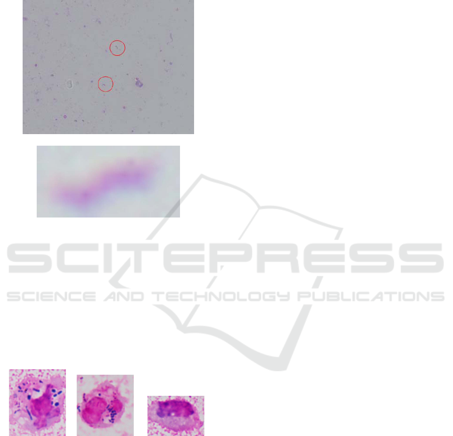
similar as dusts. Figure 1 illustrates the image con-
taining Campylobacter bacteria, which occur in just
two red circles, and a Campylobacter bacterium in
Gram stained smears images.
Figure 1: The Gram stained smears image containing
Campylobacter bacteria (upper) and a Campylobacter bac-
terium in Gram stained smears images (lower).
The difficulty of the detection of phagocytotic ac-
tivity of leukocytes is that not only phagocytotic im-
ages that a leukocyte enclose bacteria as avoiding to
its nucleus but also quasi-phagocytotic images that a
leukocyte and bacteria are just overlapping are ob-
served in Gram stained smears images. Figure 2 il-
lustrates a phagocytotic image, a quasi-phagocytotic
image and a non-phagocytotic image in Gram stained
smears images.
phagocytotic quasi-phagocytotic non-phagocytotic
Figure 2: A phagocytotic image, a quasi-phagocytotic im-
age and a non-phagocytotic image in Gram stained smears
images.
Recently, Yoshihara and Hirata (Yoshihara and
Hirata, 2021) have classified Campylobacter bacte-
ria and phagocytotic activity of leukocytes, respec-
tively, by using VGG16 (Simonyan and Zisserman,
2015) and its improvement with high accuracy. In
their work, for Campylobacter bacteria, after manu-
ally extracting the regions in which both a Campy-
lobacter bacterium occurs and no Campylobacter bac-
terium occurs, they have classified them. Also, for
phagocytotic activity of leukocytes, after manually
extracting the regions as phagocytotic images, quasi-
phagocytotic and non-phagocytoticimages, they have
classified them. Hence, the purpose of their work is
to classify Campylobacter images and phagocytotic
images which have been manually extracted, without
object detection.
On the other hand, in this paper, we detect them
with object detection. For Campylobacter bacte-
ria, after annotating the regions of Campylobacter
bacteria in training images, we detect the regions
of Campylobacter bacteria in the remained test im-
ages. For phagocytotic activity of leukocytes, after
annotating phagocytotic, quasi-phagocytoticand non-
phagocytoticregions in training images, we detect the
regions of phagocytotic activity of leukocytes in the
remained test images.
In this paper, we adopt three CNN-based object
detectors of Faster R-CNN (Ren et al., 2015), Reti-
naNet (Lin et al., 2017) and YOLOv5 (Jocher, 2020).
By using these detectors, we give experimentalresults
of detecting Campylobacter bacteria and phagocytotic
activity of leukocytes in Gram stained smears images.
1.1 Related Works
As the works dealing with Gram stained smears im-
ages, Carvajal et al. (Carvajal et al., 2014) have de-
veloped the system to learn the candidate areas from
fixed-size (51 × 38 pixels) images applicable to the
microscope test with high magnification. Hashimoto
et al. (Hashimoto et al., 2020) have developed the
system to detect Geckler classification defined by
the number of buccal squamous epithelial cells and
leukocytes for the Gram stained smears images per
100× field for the sample of sputum, in order to guar-
antee howthe Gram stained smearsimage per 1, 000×
field is quality for the microscope testing.
Lejon and Andersson (Lejon and Andersson,
2016) have developed the system to classify the bac-
teria occurring in the areas for the sample of blood
by using the template matching. Smith et al. (Smith
et al., 2018) have classified Gram negative basilli,
Gram positive cossi in clusters and Gram positive
cossi in pairs or chains from the Gram stained smear
images for the sample of blood by using CNN, after
extracting fixed size (146 × 146 pixels) images. Iida
et al. (Iida et al., 2020) have developed the system to
classify four kinds of Gram positive cocci, Gram pos-
itive bacilli, Gram negative cocci and Gram negative
bacilli from the Gram stained smear images for not
only blood but also other samples by using CNN.
Object Detection as Campylobacter Bacteria and Phagocytotic Activity of Leukocytes in Gram Stained Smears Images
535

2 OBJECT DETECTION
The purpose of this paper is to detect Campylobac-
ter bacteria and phagocytotic activity of leukocytes in
Gram stained smears images. The outline of the de-
tection is first to annotate the regions of objects as
Campylobacter bacteria and phagocytotic activity of
leukocytes in training images, and then to detect the
regions of objects in the remained test images by us-
ing the detectors.
2.1 Detectors
In this paper, we adopt the following three detectors.
Here, we implement them through PyTorch, which is
an open source machine learning library for Python.
2.1.1 Faster R-CNN
Faster R-CNN (Ren et al., 2015) is a two-stage de-
tector and consists of RPN (region proposal network)
and RoI (region of interest) pooling layer. Then,
it classifies objects by the RoI pooling layer after
proposing regions by the RPN.
To implement Faster R-CNN, we use Detec-
tron2 (Wu et al., 2019) as an object detection library,
and tune up the model of FRN+ResNeXt-101-32x8d
in PyTorch.
2.1.2 RetinaNet
RetinaNet (Lin et al., 2017) is an one-stage detec-
tor and consists of a feature pyramid network back-
bone on the top of a feedforward ResNet architec-
ture. ResNet consists of subnetworks for classifying
anchor boxes and those for regressing from anchor
boxes to ground truth object boxes. This network de-
sign is intentionally simple, which is a reason why
one-stage detectors are faster than two-stage detec-
tors.
To implement RetinaNet, we use Detectron2 (Wu
et al., 2019) as an object detection library, and tune up
the model of ResNet101 in PyTorch.
2.1.3 YOLOv5
YOLOv5 (Jocher, 2020), where YOLO is an acronym
“You only look once” and v5 means “version 5,” is
an one-stage detector integrating of the entire object
detection and classification process in a single net-
work. The network of YOLO has 24 convolutional
layers followed by 2 fully connected layers. Then,
YOLO pertains the convolutional layers on the Im-
ageNet classification task at half the resolution and
then double the resolution for detection.
To implement YOLOv5, we use a default PyTorch
library as an object detection library and tune up the
model of YOLOv5x in PyTorch.
2.2 Setting
In this paper, our computer environment is under
Google Colab that OS is Ubuntu 18.04.5 LTS, CPU
is Intel(R) Xeon(R) CPU @ 2.20GHz, RAM is 25GB
and GPU is Tesla P100-PCIE.
Also Table 1 illustrates the values of hyperparam-
eters such as epoch, batch
size and lr (learning rate).
Table 1: The values of hyperparameters that epoch,
batch
size and lr.
detector epoch batch size lr
Faster R-CNN 200 16 0.001
RetinaNet 200 16 0.001
YOLOv5 400 16 0.01
To determine the values of hyperparameters, we
tune up manually them that the values of mAP or
other AP’s are large.
2.3 Annotation
In this paper, we use 67 Gram stained smear images
for the sample of feces to detect Campylobacter bac-
teria and 101 Gram stained smear images for the sam-
ple of sputum to detect phagocytotic activity of leuko-
cytes.
Then, with helping the opinions of the medical
technologist, we annotate the regions of object as
Campylobacter bacteria and phagocytotic activity of
leukocytes in training images. For Campylobacter
bacteria, we annotate the region where an Campy-
lobacter bacteria occurs. On the other hand, for
phagocytotic activity of leukocytes, we annotate the
regions where leukocytes have phagocytotic activity
(phagocytotic), have the look of phagocytotic activ-
ity but not (quasi-phagocytotic) and have no phago-
cytotic activity (non-phagocytotic).
Figure 3 illustrates the images of annotating Cam-
phylobacter bacteria and leukocytes.
In the detection of Campylobacter bacteria (resp.,
phagocytotic activity of leukocytes), we use about
85% (resp., 90%) of images as training images includ-
ing validation images and the remained about 15%
(resp., 10%) images as test images. Since the num-
ber of training images is too small, we increase them
at triple by applying data augmentation. After anno-
tating, we resize 640×640 pixels for all the images.
ICPRAM 2022 - 11th International Conference on Pattern Recognition Applications and Methods
536
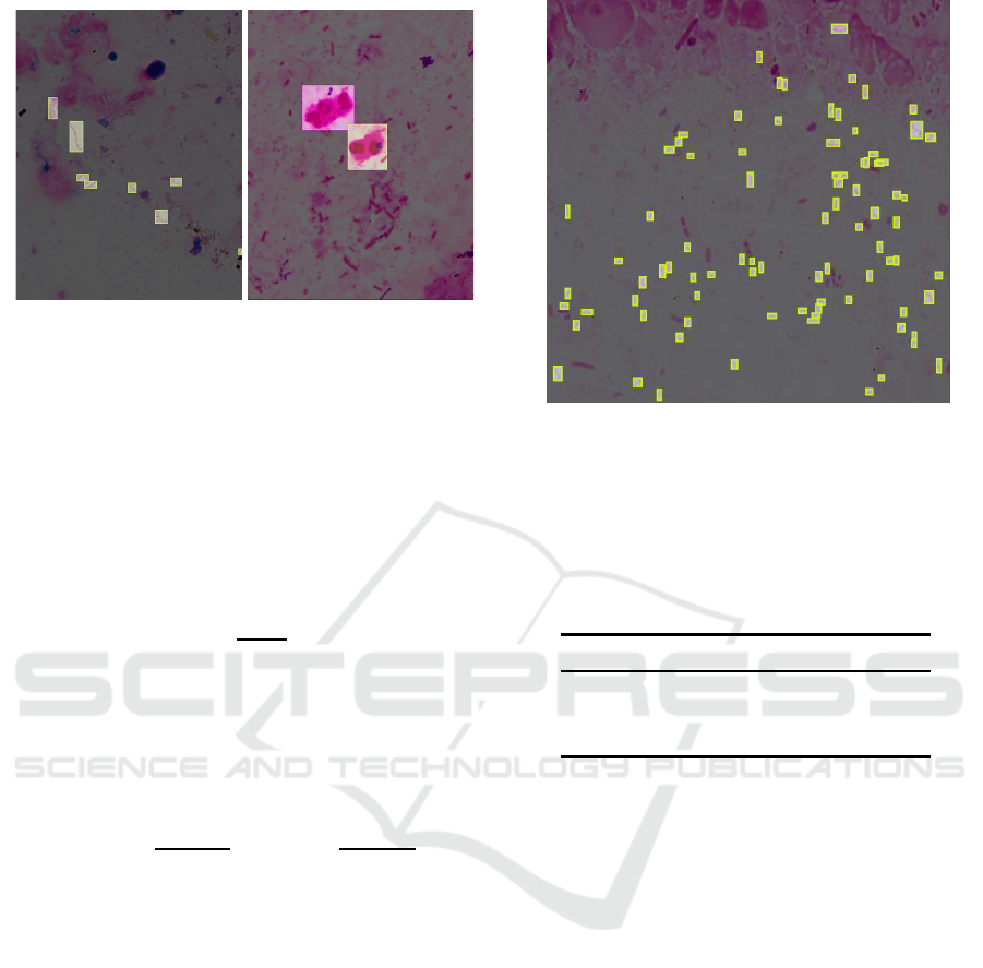
Campylobacter bacteria leukocytes
Figure 3: Annotations for Campylobacter bacteria (left) and
leukocytes (right).
2.4 Evaluation
In order to evaluate the results of the detection,
we adopt the standard measures (Everingham et al.,
2010) for object detection. First, we introduce the fol-
lowing intersection over union (IoU) between the area
P of the predicted box and the area T of the ground
truth box:
IoU =
P∩ T
P∪ T
.
For a given threshold δ (%), let TP be the number of
the predicted boxes such that IoU ≥ δ, FP the number
of the predicted boxes such that IoU < δ and FN the
number of the ground truth boxes such that IoU < δ.
Then, the standard measures of precision and recall
are defined as follows.
precision =
TP
TP+ FP
, recall =
TP
TP+ FN
.
Also, an average precision for δ (APδ) is defined as
the averagedetection precision under different recalls.
We use AP when δ = 50 and δ = 75, that is, AP50
and AP75. Furthermore, we adopt a (COCO) mean
AP (mAP) that is an average of APs when varying δ
is from 50 to 95 with a step of 5.
3 EXPERIMENTAL RESULTS
In this section, we give the experimental results to de-
tect Camylobacter bacteria and phagocytotic activity
of leukocytes.
3.1 Detection of Campylobacter
Bacteria
For the detection of Campylobacter bacteria, we use
57 training images including 10 validation images and
Figure 4: The Gram stained smear image such that Campy-
lobacter bacteria are annotated correctly.
10 test images for total 67 images. Then, Table 2 il-
lustrates the values of mAP, AP50 and AP75 to detect
Camphylobacter bacteria by using three detectors.
Table 2: The values of mAP, AP50 and AP75 to detect Cam-
phylobacter bacteria (%).
detector mAP AP50 AP75
Faster R-CNN 5.7 17.9 1.6
RetinaNet 4.1 10.5 1.6
YOLOv5 14.6 43.6 4.1
Table 2 shows that YOLOv5 has larger values of
mAP, AP50 and AP75 than Faster R-CNN and Reti-
naNet. On the other hand, for all of the Faster R-
CNN, RetinaNet and YOLOv5, the values of AP75 is
much smaller than those of mAP and AP50.
By the definition of mAP, the threshold δ such that
the value of APδ is equal to that of mAP is in the range
of [50, 75]. Then, for the detected regions of Campy-
lobacter bacteria, the overlap between the predicted
box and the ground truth box is not large as IoU.
Next, we represent the results of detecting Campy-
lobacter bacteria in Gram stained smear images. Fig-
ure 4 illustrates the Gram stained smear image such
that Campylobacter bacteria are annotated correctly.
Then, Figure 5 illustrates the result of detect-
ing Camphylobacter bacteria by Faster R-CNN, Reti-
naNet and YOLOv5 from the Gram stained smear im-
ages in Figure 4.
Figure 5 shows that both Faster R-CNN and
YOLOv5 detect many regions occurring Campy-
lobacter bacteria, whereas RetinaNet fails to detect.
By comparing the results of Faster R-CNN with
those of YOLOv5, YOLOv5 detects smaller Cam-
Object Detection as Campylobacter Bacteria and Phagocytotic Activity of Leukocytes in Gram Stained Smears Images
537
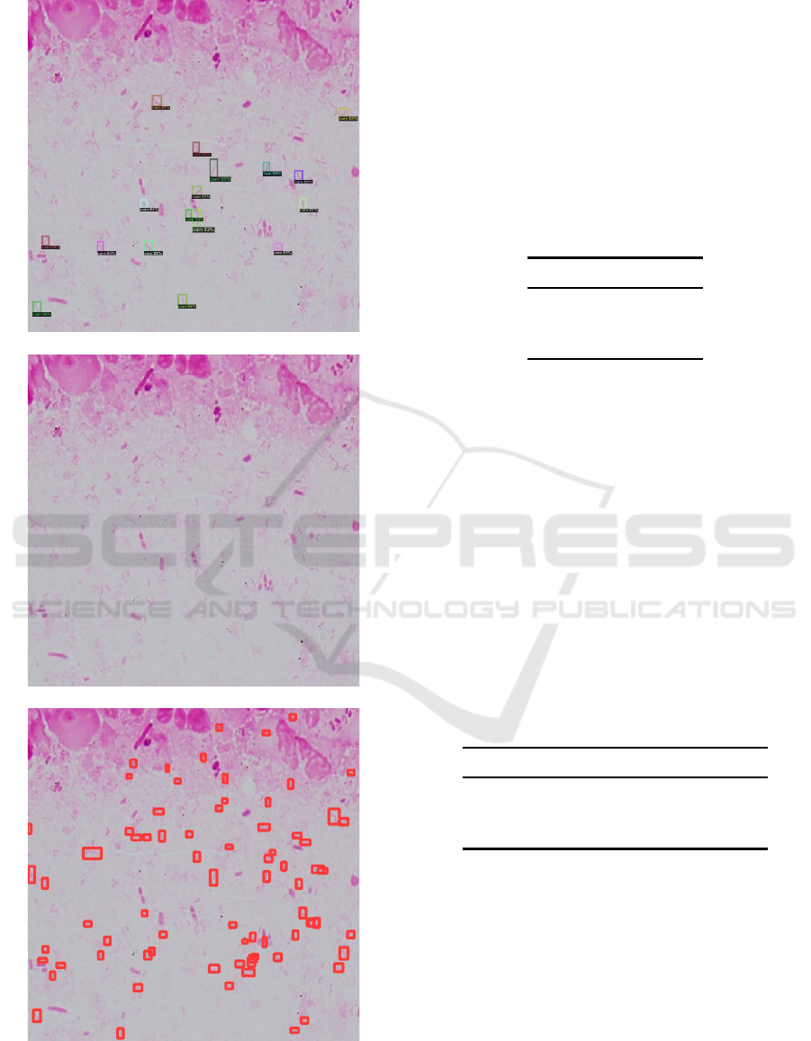
Faster R-CNN
RetinaNet
YOLOv5
Figure 5: The result of detecting Camphylobacter bacteria
by Faster R-CNN, RetinaNet and YOLOv5.
phylobacter bacteria which Faster R-CNN cannot de-
tect. Also, Faster R-CNN has the case detecting that
non-Campylobacter bacteria are Campylobacter bac-
teria. Hence, YOLOv5 is the most appropriate de-
tector. On the other hand, the value of AP50 for
YOLOv5 is 43.6%, which is the reason that many en-
twined Campylobacter bacteria exist as Figure 4.
Table 3 illustrates the average running time of de-
tectors for detecting objects of Campylobacter bacte-
ria in one image.
Table 3: The average running time (msec) for detecting ob-
jects of Campylobacter bacteria in one image.
detector time
Faster R-CNN 164.0
RetinaNet 71.8
YOLOv5 36.8
Table 3 shows that YOLOv5 is the fastest in the
three detectors, about the half of the average running
time of RetinaNet and about the quarter of that of
Faster R-CNN.
3.2 Detection of Phagocytotic Activity
of Leukocytes
For the detection of phagocytotic activity of leuko-
cytes, we use 91 training images including 10 valida-
tion images and 10 test images for total 101 images.
Then, Table 4 illustrates the values of mAP, AP50 and
AP75 to detect phagocytotic activity of leukocytes by
using three detectors.
Table 4: The values of mAP, AP50 and AP75 to detect
phagocytotic activity of leukocytes (%).
detector mAP AP50 AP75
Faster R-CNN 38.0 62.8 41.2
RetinaNet 18.4 30.0 18.8
YOLOv5 45.5 70.4 53.1
Table 4 shows that YOLOv5 has the largest values
of mAP, AP50 and AP75. In contrast to Table 2 in
Section 3.1, the values of AP75 is larger than those of
mAP in Table 4, the threshold δ such that the value of
APδ is equal to that of mAP is in the range of [75, 95].
Then, for the detected regions of phagocytotic activity
of leukocytes, the overlap between the predicted box
and the ground truth box is large as IoU.
Hence, from the viewpoint of the average pre-
cision, detecting objects of phagocytotic activity of
leukocytes is more successful than detecting objects
of Campylobacter bacteria.
ICPRAM 2022 - 11th International Conference on Pattern Recognition Applications and Methods
538
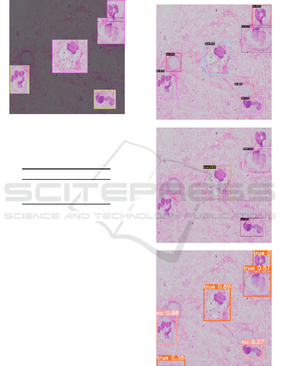
Figure 6: The Gram stained smear image such that phago-
cytotic activity are annotated correctly, where phagocytotic
and non-phagocytotic regions are enclosed by red and yel-
low colors, respectively.
Table 5: The values of AP50 for phagocytotic (pha), non-
phagocytotic (non) and quasi-phagocytotic (quasi) images
(%).
detector pha non quasi
Faster R-CNN 83.7 57.3 47.5
RetinaNet 32.1 28.1 29.7
YOLOv5 92.7 59.5 59.1
Next, we represent the results of detecting phago-
cytotic activity of leukocytes in Gram stained smear
images. Figure 6 illustrates the Gram stained smear
image such that phagocytotic activity are anno-
tated correctly, where the phagocytotic and the non-
phagocytotic regions are enclosed by red and yellow
colors, respectively.
Then, Figure 7 illustrates the result of detecting
phagocytotic activity of leukocytes by Faster R-CNN,
RetinaNet and YOLOv5 from the Gram stained smear
images in Figure 6. Here, the phagocytotic images are
labeled by “true,” the quasi-phagocytotic images by
“false” and the non-phagocytotic images by “no.”
Figure 7 shows that YOLOv5 is the most appro-
priate detector for phagocytoticactivity of leukocytes,
which detects almost leukocytes with correct classes.
Faster R-CNN detects almost leukocytes but leuko-
cytes with incorrect classes and non-leukocytes sub-
stances as leukocytes. On the other hand, RetinaNet
is insufficient to detect leukocytes.
Table 5 illustrates the values of AP50 for phago-
cytotic, non-phagocytotic and quasi-phagocytotic im-
ages.
Table 5 shows that the value of AP50 for phagocy-
totic images by YOLOv5 is much larger than those by
Faster R-CNN
RetinaNet
YOLOv5
Figure 7: The result of detecting phagocytotic activity of
leukocytes by Faster R-CNN, RetinaNet and YOLOv5.
Object Detection as Campylobacter Bacteria and Phagocytotic Activity of Leukocytes in Gram Stained Smears Images
539
Faster R-CNN and RetinaNet. Since the purpose of
this paper is to detect phagocytotic images correctly,
YOLOv5 is the most appropriate detector of the pur-
pose. Also, the values of AP50 for non-phagocytotic
images by all the detectors are very small. The rea-
son is that quasi-phagocytotic images is misclassified
to non-phagocytotic images. Nevertheless, if we re-
gard both quasi- and non-phagocytotic images as non-
phagocytotic images, this misclassification can be ig-
nored to detect phagocytotic images correctly. As a
result, YOLOv5 succeeds to detect phagocytotic im-
ages correctly.
Table 6 illustrates the average running time of de-
tectors for detecting objects phagocytotic activity of
Leukocytes in one image.
Table 6: The average running time (msec) for detecting ob-
jects of phagocytotic activity of leukocytes in one image.
detector time
Faster R-CNN 146.0
RetinaNet 69.6
YOLOv5 34.9
Table 6 shows that YOLOv5 is the fastest in the
three detectors, about the half of the average run-
ning time of RetinaNet and about the quarter of that
of Faster R-CNN. By incorporating Table 3 in Sec-
tion 3.1 with Table 6, we can conclude that YOLOv5
is the fastest in the three detectors for not only
Campylobacter bacteria but also phagocytotic activ-
ity of Leukocytes.
Furthermore, the running time of detecting objects
of Camphylobacter bacteria is slightly larger than that
of phagocytotic activity of leukocytes but they are al-
most equal. Hence, the running time of the object
detection in this paper is independent from the object
as the target.
4 CONCLUSION AND FUTURE
WORKS
In this paper, we have detected Campylobacter bac-
teria and phagocytotic activity of leukocytes in Gram
stained smear images by using the detectors of Faster
R-CNN, RetinaNet and YOLOv5. Then, RetinaNet
have failed to detect them, and YOLOv5 is more ap-
propriate to detect them than Faster R-CNN.
In particular, for phagocytotic activity of leuko-
cytes, YOLOv5 have succeeded to detect almost
leukocytes with correct classes. On the other hand,
YOLOv5 have succeeded to detect Campylobac-
ter bacteria in many cases, whereas the cases that
Campylobacter bacteria have not detected exist. The
reason is that YOLOv5 does not work well for the im-
ages having many small objects.
Then, it is a future work to improve YOLOv5 to
work well for such images. In particular, we apply
YOLOv5 after decreasing the number of objects by
dividing an image and enlarging the objects.
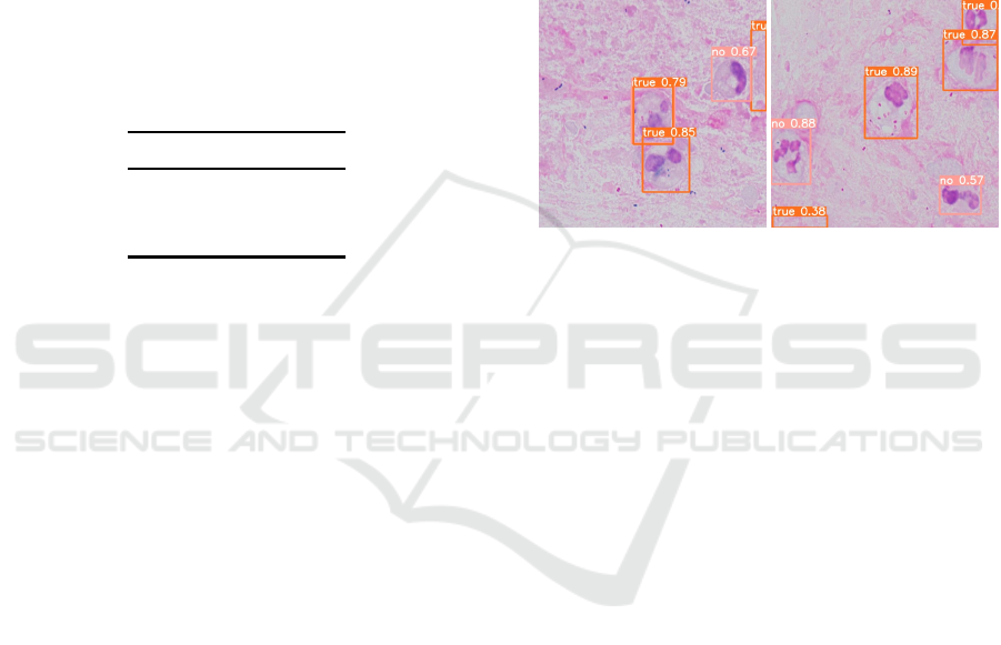
Whereas YOLOv5 succeed to detect phagocytotic
images at 90% under AP50 in Table 5 as stated
in Section 3.2, there exist some images that non-
phagocytotic images are detected as phagocytotic.
Figure 8 illustrates such images.
Figure 8: The images that non-phagocytotic images are de-
tected as phagocytotic.
The upper-right region labeled by “no” in the left
image in Figure 8 and the lower-left and lower-right
regions labeled by “no” in the right image in Figure 8
are not leukocytes. Even if recall is more important
than precision in the medical data, it is a future work
to solve this misclassification by improving annota-
tions in test images.
Since the number of training images in this paper
is too small to succeed object detection, it is neces-
sary to collect the large number of training images by
the medical technologist. Also since we use Gram
stained smears images photographed under the same
environment, it is necessary to collect the images un-
der the several environment, by using different equip-
ment and differentbrightness. These are future works.
REFERENCES
Bartholomew, J. and Mittwer, T. (1952). The Gram stain.
Bacteriol. Rev., 16:1–29.
Carvajal, J., Smith, D., Zhao, K., Wiliem, A., Finucane, P.,
Hobson, P., Jennings, A., McDougall, R., and Lovell,
B. (2014). An early experience toward developing
computer aided diagnosis for Gram-stained smear im-
ages. In Proc. CVPR’14, pages 62–28.
Everingham, M., Gool, L. V., Williams, C. K. I., Winn, J.,
and Zisserman, A. (2010). The PASCAL visual object
classes (VOC) challenge. Internat. J. Comput. Vision,
88:303–338.
ICPRAM 2022 - 11th International Conference on Pattern Recognition Applications and Methods
540

Hashimoto, K., Iida, R., Hirata, K., Matsuoka, K., and
Yokoyama, S. (2020). Detecting Geckler classifica-
tion from Gram stained smears images for sputum. In
Proc. ICPRAM’20, pages 469–476.
Iida, R., Hashimoto, K., Hirata, K., Matsuoka, K., and
Yokoyama, S. (2020). Detection system of Gram
types for bacteria from Gram stained smears images.
In Proc. ICPRAM’20, pages 477–484.
Jocher, G. (2020). YOLOv5. https://github.com/ultralytics/
yolov5.
Lejon, S. and Andersson, E. (2016). Semi-automatic
segmentation, detection and classification of Gram
stained bacteria in blood sample. Master Thesis,
Lund University.
Lin, T., Goyal, P., Girshick, R., He, K., and Dollar, P.
(2017). Focal loss for dense object detection. In Proc.
ICCV’17, pages 2999–3007.
Ren, S., He, K., Girshick, R., and Sun, J. (2015). Faster R-
CNN: Towards real-time object detection with region
proposal networks. In Proc. NIPS’15, pages 91–99.
Simonyan, K. and Zisserman, A. (2015). Very deep convo-
lution networks for large-scale image recognition. In
Proc. ICLR’15.
Smith, K., Kang, A., and Kirby, J. (2018). Automated in-
terpretation of blood culture Gram stains by use of a
deep convolutional neural network. J. Clin. Microbio.,
56:e01521–17.
Wu, Y., Kirillov, A., Massa, F., Lo, W.-Y., and Gir-
shick, R. (2019). Detectron2. https://github.com/
facebookresearch/detectron2/.
Yoshihara, K. and Hirata, K. (2021). Detecting Camphy-
lobacter bacteria and phagocytotic activity of leuko-
cytes from Gram stained smear images. In Proc.
ESKM’21, pages 10–15.
Object Detection as Campylobacter Bacteria and Phagocytotic Activity of Leukocytes in Gram Stained Smears Images
541
