
U-Net-based DFU Tissue Segmentation and Registration on Uncontrolled
Dermoscopic Images
Yanexis Toledo
1 a
, Leandro A. F. Fernandes
1 b
, Silena Herold-Garcia
2 c
and Alexis P. Quesada
3
1
Instituto de Computac¸
˜
ao, Universidade Federal Fluminense, Niter
´
oi, Brazil
2
Facultad de Matem
´
atica y Computaci
´
on, Universidad de Oriente, Santiago de Cuba, Cuba
3
Hospital General Dr. Juan Bruno Zayas Alfonso, Santiago de Cuba, Cuba
Keywords:
Semantic Segmentation, Uncontrolled Viewpoint, Epipolar Constraints, Image Registration.
Abstract:
Diabetic Foot Ulcers (DFUs) are aggressive wounds with high morbimortality due to their slow healing ca-
pacity and rapid tissue degeneration, which cause complications such as infection, gangrene, and amputation.
The automatic analysis of the evolution of tissues associated with DFU allows the quick identification and
treatment of possible complications. In this paper, our contribution is twofold. First, we present a new DFU
dataset composed of 222 images labeled by specialists. The images followed the healing process of patients
of an experimental treatment and were captured under uncontrolled viewpoint and illumination conditions. To
the best of our knowledge, this is the first DFU dataset whose images include the identification of background
and six different classes of tissues. The second contribution is an U-Net-based segmentation and registration
procedure that uses features computed by hidden layers of the network and epipolar constraints to identify
pixelwise correspondences between images of the same patient at different healing stages.
1 INTRODUCTION
During the treatment of Diabetic Foot Ulcers (DFUs),
the patient’s foot undergoes significant transforma-
tions. The different tissues alert the specialists about
the clinical evolution of the patient’s condition. Typ-
ically, the analysis of these ulcers is carried out vi-
sually by specialists, which is a process prone to er-
rors. The automatic analysis of the evolution of DFUs
can mitigate the problems arising from manual in-
spection. Two processes are required as part of the
automatic analysis: the segmentation of the image re-
gions corresponding to different tissues and the reg-
istration of the images of each patient throughout the
treatment. In this paper, we address both processes.
The semantic segmentation of DFU images requires
robust computer vision techniques in the face of low
contrast and variations on the image acquisition con-
ditions. Deep learning models are state-of-the-art in
semantic segmentation (Hao et al., 2020). However,
the success of these models depends on the quality
of training datasets. Unfortunately, large sets of DFU
a
https://orcid.org/0000-0003-4103-0225
b
https://orcid.org/0000-0001-8491-793X
c
https://orcid.org/0000-0001-9238-3472
images are not available, and the existing ones label
image pixels as wound and non-wound only. Our
first challenge in this work was to handle these is-
sues. In our approach, we have used images from the
Foot Ulcer Segmentation Challenge 2021 (FUSeg)
and the Medetec Wound Database to train an initial
version of our U-Net-based segmentation model con-
sidering the two usual classes. Segmentation consid-
ering six different classes of tissue, plus background,
was obtained by transfer learning from the 2-class
to the 7-class model trained with a new DFU image
dataset built for this work (Figure 1). Our dataset
includes images captured under uncontrolled condi-
tions from patients undergoing an experimental treat-
ment in the General Dr. Juan Bruno Zayas Alfonso
Hospital in Cuba. Each image in our dataset has a
mask that labels the pixels as background, epithelial-
ization, healthy skin, granulation tissue, slough tis-
sue, necrotic tissue, and exposed tendon.
The second challenge in this work was to de-
velop an image registration technique to keep track
of wound evolution along with the treatment. In con-
trast to DFU analysis techniques described in the lit-
erature (Sol
´
ıs-S
´
anchez et al., 2016), which consider
highly controlled environments during image acqui-
sition, in this work, we assume that hard capturing
510
Toledo, Y., Fernandes, L., Herold-Garcia, S. and Quesada, A.
U-Net-based DFU Tissue Segmentation and Registration on Uncontrolled Dermoscopic Images.
DOI: 10.5220/0010868600003124
In Proceedings of the 17th International Joint Conference on Computer Vision, Imaging and Computer Graphics Theory and Applications (VISIGRAPP 2022) - Volume 4: VISAPP, pages
510-517
ISBN: 978-989-758-555-5; ISSN: 2184-4321
Copyright
c
2022 by SCITEPRESS – Science and Technology Publications, Lda. All rights reserved
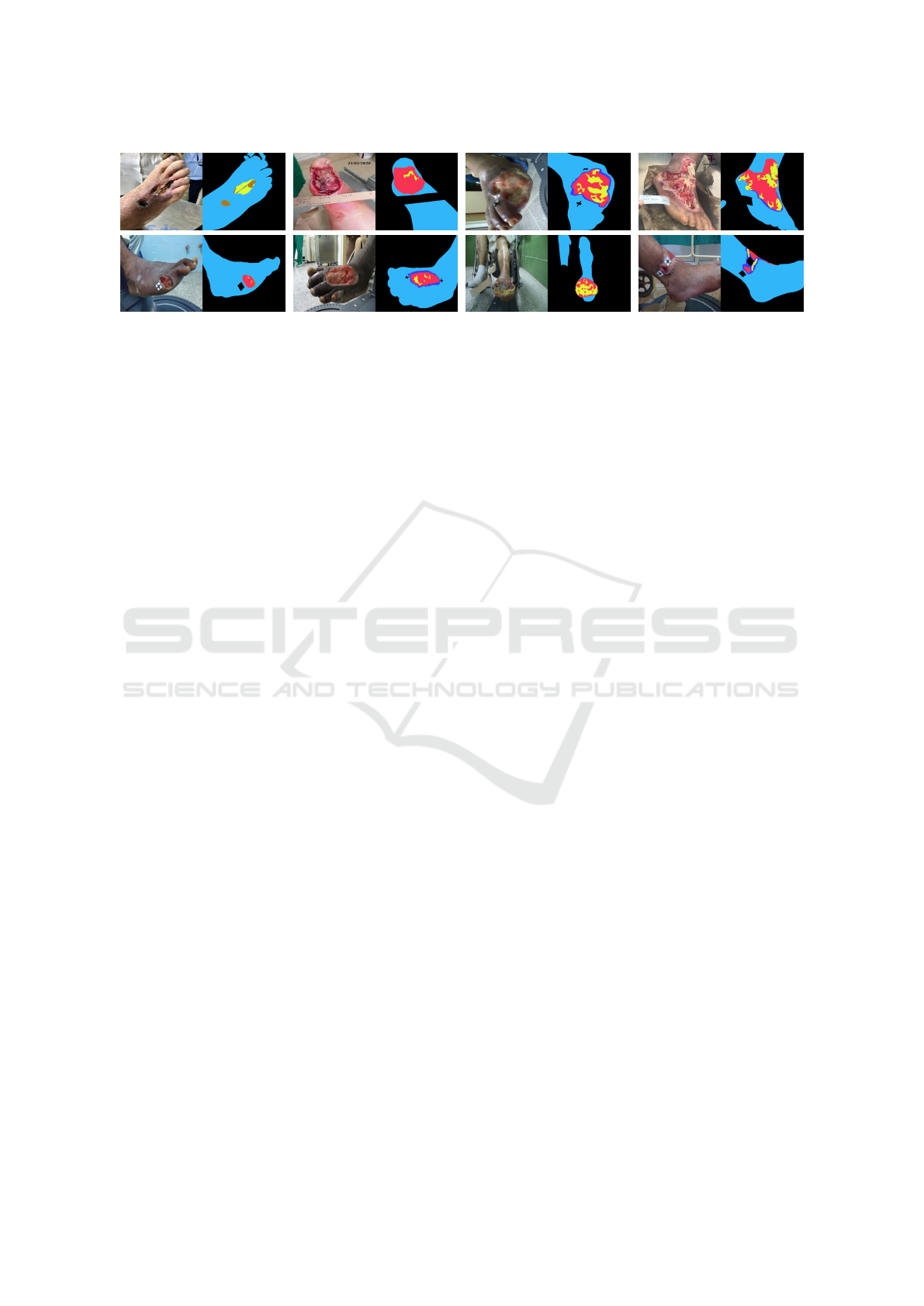
Figure 1: Images from our DFU dataset and their respective segmentation masks. The mask colors correspond to back-
ground , epithelialization , healthy skin , granulation tissue , slough tissue , necrotic tissue , and exposed tendon .
restrictions may prevent the application of an analysis
technique in realistic scenarios. For such, the regis-
tration process must be robust to the temporal evo-
lution of the wound’s appearance, variations on dis-
tance, point of view, and lighting conditions. We use
feature maps computed by the semantic segmentation
model and its segmentation results to extract robust
descriptors for tiles in the wounds and health skin re-
gions. In turn, our registration approach uses epipo-
lar constraints (Hartley and Zissermann, 2004) to es-
tablish correspondences and reduce the occurrence of
matching outliers between pairs of images of the same
patient acquired on different dates.
Our main contributions can be summarized as:
(i) a new DFU dataset composed of 222 images
whose pixels were labeled by specialists considering
six types of tissues and background; and (ii) a new
U-Net-based segmentation and registration procedure
for DFU images. To the best of our knowledge, our
DFU dataset is the one including the highest number
of labels in the annotation masks. Also, our registra-
tion approach is the first to use features extracted from
an U-Net as image descriptors for image registration.
2 RELATED WORK
DFU Image Segmentation. Convolutional Neural
Networks (CNNs) are cutting edge in the image seg-
mentation task of complex wounds such as DFUs.
Although their performance varies according to the
training dataset, they all show promising results for
the 2-class segmentation problem (wound and non-
wound) (Goyal et al., 2017; Wang et al., 2020; Wagh
et al., 2020), and few address 4 classes (external skin,
necrosis, granulation, and slough) (Kaswan et al.,
2020). The main advantages of the U-Net architec-
ture are the reduced times necessary for training and
the good performance even with few training sam-
ples. The higher convergence speed of U-Nets during
the training phase is related to the jump connections
between the encoder and the decoder blocks, which
contribute to smooth the descent path of the gradient
towards the global minimum.
Image Registration. Klein et al. (2009) perform
elastic alignment of intensity medical image via the
estimation of displacement vectors. Klein’s et al. ap-
proach is capable of representing complex local dis-
tortions but is prone to compatibility issues and re-
quires excessive execution times. By combining non-
elastic and elastic alignments, Zhang et al. (2019)
are capable of performing natural (non-medical) im-
age registration with the robustness of the parametric
alignment. Experimental results show that Zhang’s et
al. method is accurate and surpasses a selection of
last-generation image alignment approaches. Unfor-
tunately, the implementation is not available, which
prevents its use and continuity by other researchers.
Long et al. (2014) proposed the applicability of fea-
ture vectors extracted from CNNs in tasks such as se-
mantic segmentation and robust correspondence esti-
mation. Recent works have attempted to analyze and
explain this overwhelming success (Dara and Tumma,
2018). Our work extends these studies to DFU images
using features computed in a segmentation model.
Current techniques that analyze the evolution of
wounds consider controlled environments (Sol
´
ıs-
S
´
anchez et al., 2016). The absence of restrictions in
the image capture process shows that our wound reg-
istration technique goes beyond state of the art ap-
proaches.
3 MATERIALS AND METHODS
Our approach uses an U-Net CNN architecture (Ron-
neberger et al., 2015) to segment DFU images con-
sidering six different types of tissues, plus the back-
ground (Figure 2, top), and to extract robust descrip-
tors for efficient region matching and subsequent im-
age registration (Figure 2, bottom).
U-Net-based DFU Tissue Segmentation and Registration on Uncontrolled Dermoscopic Images
511
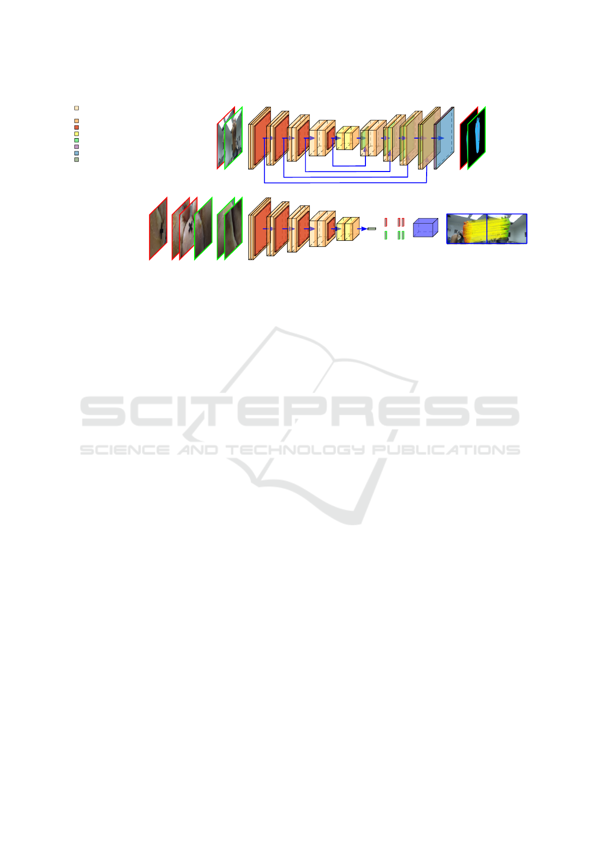
64
64
Down1
128
128
Down2
256
256
Down3
512
512
Down4
512
512
Junction
512
256
Up1
256
128
Up2
128
64
Up3
64
64
Up4
7
Out
Resized
Input Images
Resulting
Segmentation
... ...
ROI Tiles of the
Input Images
64
64
Down1
128
128
Down2
256
256
Down3
512
512
Down4
512
512
Junction
512
Peek
Conv2d(in
k
, out
k
, 3x3, bias, stride=1, padding=1)
ReLU
MaxPool2d(2x2, stride=2)
Conv2d(512, 512, 3x3, bias, stride=1, padding=1)
Softmax
Conv2d(64, 7, 1x1, bias, stride=1)
CentralPeek
...
...
Descriptors
Matching
Procedure
Resulting
Registration
3
3
UpSample(scale=2, bilinear)
+ BatchNorm2d(out
k
, momentum=0.1)
3
3
3
3
3
3
Figure 2: Overview of the proposed segmentation (top) and registration (bottom) methods.
3.1 Our Diabetic Foot Ulcer Dataset
Our dataset was collected at a hospital center over one
year, between 2019 and 2020, by specialists in vas-
cular wounds who carried out an experimental study
on DFUs. The dataset is composed of 222 anno-
tated DFU images of 28 patients. The image his-
tory of each patient consists of weekly image cap-
tures from the first time the patient attended the treat-
ment until the wound is completely closed, with an
average of 9 captures per subject. The images were
taken by four different digital camera models un-
der uncontrolled illumination conditions, with vari-
ous viewpoints and backgrounds. Image resolution
ranges from 522 × 692 to 4608 × 3456 pixels. The
characteristics of the patients are pretty diverse, more-
over, there is an evident imbalance among the types
of tissues observed in the images. Necrotic tissue and
exposed tendon are rare, while most images contain
granulation tissue, slough tissue, and epithelialization
(Figure 1). The segmentation masks in our dataset
were created by an specialist using the free online
Computer Vision Annotation Tool. All skin informa-
tion within the image, whether or not it was from the
patient, was marked as healthy skin tissue, and inter-
fering objects such as paper markers, among others,
were annotated as background.
3.2 Segmentation Network
The network expects as input a batch of RGB images
having 224 × 224 pixels and produces one tensor with
224 × 224 pixels and seven channels for each image
in the batch. Images with a resolution different than
expected must be resized to fit the input shape, and
the result is resized back to the original image resolu-
tion. The channels of the output encode the probabil-
ities of a given pixel be related to the classes consid-
ered in our dataset. The segmentation network has a
4-block encoder-decoder structure. From the first to
the last encoder block DownK , the number of output
channels in the convolutions is 64, 128, 256, and 512,
respectively. The encoder blocks are followed by the
Junction block. These five blocks are in charge of
extracting the essential characteristics of the input im-
ages, which will be used here for segmentation and in
Section 3.3 as descriptors for image registration. The
decoder blocks UpK in Figure 2 recover the resolution
of the input image. The result of each up-sample in
these blocks is concatenated to the feature map pro-
duced by the encoder layer of its same level. Thus,
the number of input channels of the first convolution
in each UpK block is, respectively, 1024, 512, 256,
and 128. The Out block returns per-pixel probability
distributions of the seven classes.
3.3 Image Registration Procedure
We restrict the registration process to the portions of
the input image classified as non-background. Ac-
cording to our experience, classic algorithms for au-
tomatic detection of features such as SIFT and SURF
fail on describing health skin and wound textures for
three main reasons: (i) the keypoints are usually at-
tracted by the boundaries of the foot; (ii) health skin
regions are poor in texture; and (iii) the appearance of
the wounded regions changes over time. In contrast,
it has been shown that features computed by the im-
age classification networks such as VGG and ResNet
are successful as a source of image descriptors (Long
et al., 2014; Dara and Tumma, 2018). We use fea-
ture vectors computed by the Junction block of the
U-Net as source of region descriptors, even though
the amount of DFU images available for training is
much smaller than the number of images in datasets
for image classification, like CIFAR-10 and COCO.
VISAPP 2022 - 17th International Conference on Computer Vision Theory and Applications
512
The steps of our registration procedure are feature ex-
traction, feature matching, and epipolar pruning with
dense registration.
Feature Extraction. We split the (original) input
image into overlapping tiles having the size expected
by the first layer of the U-Net (i.e., 224 × 224 pixels)
and stride s. In our experiments, s was empirically
set to 10 pixels after analyzing its impact on process-
ing time. The tiles whose central pixel were classified
as one of the six types of tissue during the segmenta-
tion process (Figure 2, top) are submitted to the en-
coder blocks of the U-Net (Figure 2, bottom), and the
feature vector at the center of the 14 × 14 × 512 map
produced by the Junction block is taken as the de-
scriptor for a given tile (Peek operation). Thus, for
each tile we have a feature vector d
p
associated to the
image location p at tile’s center.
Feature Matching. After extracting sets of descrip-
tors from two images acquired from the same patient,
the feature matching step normalizes the feature vec-
tors to unit vectors using the L2 norm and exhaus-
tively computes the Sum of Squared Distances (SSD)
between the normalized features from each set. For
each normalized entry of the first set, the procedure
returns the closest one in the second set if the SSD
distance is less than the match threshold of 1% from
a perfect match. Due to normalization, the SSD val-
ues range is [0, 4]. Multiple features in the first set
can match one feature in the second one. The feature
matching step returns pairs of correspondent sparse
image locations p ↔ p
0
, where p belongs to a non-
background region of image I and p
0
is in a non-
background region of image I
0
.
Epipolar Pruning with Final Dense Registration.
We use the correspondences p ↔ p
0
to estimate the
epipolar geometry of the cameras used to capture the
DFU images. For that, we apply RANSAC, which in
addition removes most ambiguities, as it keeps track
of the set of inliers related to the estimated funda-
mental matrix F, but that nevertheless also usually
contains false-positive matches. RANSAC alone can-
not perform elastic (non-linear) dense image regis-
tration from a sparse set of keypoints. Algorithm 1
describes how we use the RANSAC’s consensus set,
epipolar constraints, and angular pondering to per-
form dense elastic registration between DFU images
I and I
0
. As a result of the image registration, one
must expect coherence on displacement vectors com-
puted from the location of a point x ∈ I to its cor-
responding point x
0
∈ I
0
(see Figure 4, right). The
same applies to the keypoints in the subset of inliers
Algorithm 1: Epipolar Pruning with Final Dense Registra-
tion.
Data: Sparse set of correspondences p ↔ p
0
of keypoints from images I and I
0
of
the same patient
Result: Dense elastic registration of the
non-background regions of the input
1 Use RANSAC to estimate the matrix F that
best fit the input set of correspondences and
the respective subset of inliers
2 Compute the angles between the x-axis and
displacement vectors
~
v = p
0
− p using
p ↔ p
0
pairs from the subset of inliers
3 Compute the normalized cumulative
histogram of angles and call it the CDF of
angular values
4 for each pixel location x segmented as
non-background in image I do
5 Use the epipolar constraint to compute
the epipolar line l
0
= F x in image I
0
6 for each pixel x
0
∈ l
0
and in a
non-background region of I
0
do
7 Compute and store the weighted
distance
1
p
x,x
0
k
d
x
− d
x
0
k
2
L2
within x
(use the CDF to compute p
x,x
0
)
8 end
9 Take the smallest distance and set the
respective point x
0
as correspondent to x
10 end
11 The set of x ↔ x
0
pairs define to the dense
elastic registration of I and I
0
returned by RANSAC. Thus, after determining the
fundamental matrix F (Algorithm 1, line 1), we use
the consensus set to compute the cumulative distri-
bution functions (CDF) of the angles between the x-
axis and the displacement vectors
~
v = p
0
− p of the
p ↔ p
0
pairs in the subset of inliers (lines 2 and 3).
We have assumed a discrete set of 360 angular val-
ues to compute the CDF. Next, for each pixel lo-
cation x = (x,y, 1)
|
within a non-background region
of I , we look for its correspondent pixel location
x
0
= (x
0
,y
0
,1)
|
in image I
0
(lines 4 to 10). We use the
epipolar constraint given by l
0
= F x to compute the
epipolar line l
0
= (A
0
,B
0
,C
0
)
|
(line 5), and the Bresen-
ham algorithm to traverse the portions of l
0
that inter-
sect with a non-background region in I
0
(lines 6 to 8).
A, B, and C are the coefficients of the general form of
the equation of a straight line, i.e., Ax + By +Cw = 0.
Recall that we extracted feature vectors for tiles in
both images. Here, we assume that pixels x and x
0
are related to the feature vectors d
p
and d
p
0
com-
puted at the tile center closest to x and x
0
, respec-
U-Net-based DFU Tissue Segmentation and Registration on Uncontrolled Dermoscopic Images
513

tively. Rather than simply comparing the features as-
sociated with x and x
0
, we use the inverse angle prob-
abilities as a weighting factor of the squared distance
of the descriptors while traversing the epipolar lines
(line 7). Formally, for a given pixel location x, we
retrieve x
0
= argmin
x
0
∈l
0
1
p
x,x
0
k
d
x
− d
x
0
k
2
L2
as its corre-
sponding pixel location, where p
x,x
0
is the probabil-
ity estimated for the angle between the x-axis and the
displacement vector from x to x
0
. The use of epipo-
lar constraints enhanced with angular restrictions is
an original idea of our work. Our experience shows
that the presence of outliers is mitigated when
1
p
x,x
0
is
used to weight the distance between features.
4 EXPERIMENTS AND RESULTS
We have implemented the network (Section 3.2) us-
ing PyTorch 1.4 and the image registration proce-
dure (Section 3.3) using MATLAB R2020a
1
. We
ran our experiments in an Intel Xeon E5-2698 v4
CPU with 2.2Ghz, 512Gb of RAM, and 8 GPUs
NVIDIA Tesla P100-SXM2 with 16Gb of memory
each. Section 4.1 presents experiments tailored to
compare DFU segmentation results produced by our
U-Net against ENet (Paszke et al., 2016), Deeplab V3
with ResNet-50 backbone (Chen et al., 2017), and
SegNet (Badrinarayanan et al., 2017). Since we don’t
have enough images to train models that consider 7
classes from scratch, we first initialized the parame-
ters of the networks with random values and trained
the models for the wound and non-wound classes.
Then, we applied transfer learning and performed
the second phase of the training process using the 7-
class dataset. Our experiments demonstrate the influ-
ence of data augmentation and transfer learning in the
segmentation model’s quality. Section 4.2 presents
the results of the automatic registration procedure for
pairs of DFU images from the same patient captured
at different weeks. We take advantage of the segmen-
tation model obtained in Section 4.1 to extract sparse
feature descriptors from DFU images regions.
4.1 Segmentation Results
Data Preparation. We built the dataset used to
train binary segmentation models by joining the
FUSeg Dataset (Wang et al., 2021) and the Mede-
tec Wound Database (Medetec, 2021). Altogether,
this 2-class dataset includes 1,919 images. For the
7-class segmentation problem, the dataset used in
our experiments includes the 222 images described
1
https://github.com/Prograf-UFF/DFU
in Section 3.1, plus 35 DFU images from the Mede-
tec Wound Database not included in the binary seg-
mentation dataset. These extra images were also an-
notated by a specialist. We took each set of images
and randomly split them into training (60%), valida-
tion (20%), and test (20%) subsets. Then, the train-
ing subsets were augmented with images produced by
the Albumentations library considering nine random
transformations: Gaussian blur, motion blur, optical
distortion, brightness contrast, scale, translation, ro-
tation, and horizontal and vertical flip. In the end, the
training subsets of the 2- and 7-class datasets included
5,765 and 1,550 images, respectively.
Hyperparameter Tuning. We performed hyperpa-
rameter tuning in both phases of the training process
of the networks via a Bayesian approach (Bergstra
et al., 2013), computing the F1-Score and the Cross
Entropy (CE) as metrics for validation in, respec-
tively, the 2- and 7-class segmentation tasks. Also,
we used Hyperband (Li et al., 2017) as stopping cri-
teria, with min iter = 20 and η = 3, and a limit of
30 runs per sweep due to time constraints. The im-
plementations of Bergstra et al. (2013) and Li et al.
(2017) procedures are available in the Weights & Bi-
ases toolset. The hyperparameters considered were:
batch size (from 2 to 20), learning rate (from 10
−7
to 10
−1
), and optimizer type (Adam, SGD, and RM-
Sprop). Tables 1 and 2 show the hyperparameter val-
ues of the best models found in each scenario. The
final scores were calculated on the test subset.
Discussion. DeepLab models have better average
performance in 2-class (Table 1) and 7-class seg-
mentation tasks (Table 2), closely followed by U-Net
models. However, it is important to comment that we
have observed advantages in using U-Net to meet the
purpose of our research (i.e., segmentation and regis-
tration of DFU images). The first advantage is that
they require less training time and significantly less
GPU memory than DeepLab models. The second ad-
vantage is that the features extracted by U-Net, which
are also used to register pairs of images, are vectors
with only 512 components, while the latent features
produced by DeepLab have 2,048 components. We
have observed that using descriptors with four times
more elements does not improve the quality of the
registration process of DFU images but dramatically
impacts its computational cost. In general, the perfor-
mance of ENet and SegNet in DFU image segmen-
tation tasks is much lower than U-Net and DeepLab,
specially on the 7-classes case. In Table 2, the dif-
ference between mean F1-Score and mean Accuracy
(Acc) of SegNet to U-Net reaches 0.15, and the dif-
VISAPP 2022 - 17th International Conference on Computer Vision Theory and Applications
514
Table 1: Hyperparameters, training values, and scores of 2-class segmentation models trained on different scenarios of data
augmentation (DA). Bold and underlined values highlight the best and second-best results among the compared models.
DA Network
Hyperparameters Training Scores
Batch
Size
Learning
Rate
Optimizer Epochs Runtime
F1
IoU Acc Recall
Val. Test
–
U-Net 6 0.0486 SGD 48 0:35:03 0.82 0.89 0.90 1.00 0.88
ENet 20 0.0016 Adam 56 0:12:27 0.79 0.86 0.87 0.99 0.84
DeepLab 5 0.0586 SGD 36 2:02:14 0.87 0.90 0.91 1.00 0.92
SegNet 3 0.0554 Adam 36 0:33:20 0.50 0.72 0.78 0.99 0.59
X
U-Net 6 0.0853 SGD 66 2:43:54 0.86 0.91 0.91 1.00 0.89
ENet 5 0.0323 SGD 87 3:04:01 0.85 0.89 0.90 1.00 0.88
DeepLab 8 0.0643 SGD 20 5:49:51 0.85 0.91 0.92 1.00 0.93
SegNet 10 0.0116 SGD 67 3:48:44 0.76 0.86 0.87 0.99 0.79
Table 2: Hyperparameters, training values, and scores of 7-class segmentation models on different scenarios of DA and TL.
TL DA Network
Hyperparameters Training Scores
Batch
Size
Learning
Rate
Optimizer Epochs Runtime
CE
Mean
F1
Mean
IoU
Mean
Acc
Mean
Recall
Val. Test
– –
U-Net 12 0.0228 SGD 41 0:12:26 0.28 0.31 0.91 0.40 0.91 0.46
ENet 4 0.0041 RMSprop 56 0:18:08 0.40 0.38 0.88 0.29 0.88 0.33
DeepLab 3 0.0142 SGD 26 0:09:25 0.18 0.20 0.94 0.45 0.94 0.53
SegNet 3 0.0051 RMSprop 58 0:09:45 0.50 0.48 0.82 0.22 0.82 0.27
– X
U-Net 11 0.0105 SGD 41 0:31:17 0.18 0.21 0.94 0.46 0.94 0.55
ENet 20 0.0286 Adam 57 0:24:01 0.20 0.23 0.93 0.41 0.93 0.47
DeepLab 7 0.0534 SGD 24 1:48:58 0.14 0.15 0.95 0.49 0.95 0.57
SegNet 11 0.0223 SGD 54 0:36:25 0.34 0.34 0.89 0.34 0.89 0.42
X –
U-Net 7 0.0853 SGD 44 0:09:05 0.23 0.24 0.92 0.38 0.92 0.44
ENet 13 0.0521 SGD 52 0:04:11 0.27 0.31 0.91 0.32 0.91 0.38
DeepLab 4 0.0484 SGD 24 0:09:46 0.21 0.21 0.94 0.38 0.94 0.44
SegNet 7 0.0941 SGD 26 0:03:15 0.59 0.68 0.77 0.17 0.77 0.21
X X
U-Net 9 0.0890 SGD 40 0:38:30 0.17 0.19 0.94 0.48 0.94 0.55
ENet 6 0.0871 SGD 51 0:54:00 0.20 0.23 0.93 0.39 0.93 0.45
DeepLab 7 0.0549 SGD 30 1:27:50 0.14 0.17 0.95 0.48 0.95 0.56
SegNet 7 0.0445 SGD 39 0:48:03 0.47 0.44 0.85 0.32 0.85 0.39
Table 3: Per class metric scores of U-Net models trained for 7 classes on different scenarios of transfer learning (TL) and data
augmentation (DA). Color/Class legend in Figure 1.
TL DA
F1 IoU
– – 0.96 0.00 0.88 0.71 0.46 0.40 0.00 0.92 0.00 0.78 0.55 0.30 0.25 0.00
– X 0.98 0.21 0.92 0.77 0.57 0.46 0.00 0.95 0.11 0.85 0.63 0.39 0.30 0.00
X – 0.96 0.03 0.89 0.75 0.48 0.05 0.00 0.93 0.02 0.80 0.60 0.31 0.03 0.00
X X 0.98 0.26 0.93 0.81 0.54 0.47 0.00 0.96 0.15 0.87 0.68 0.37 0.31 0.00
ference between mean Intersection over Union (IoU)
and mean Recall of the same architectures are up to
0.21 and 0.23, respectively. For ENet models, the
higher differences in the mean scores concerning U-
Net range from 0.03 (F1 and Acc) to 0.13 (Recall).
According to our experiments on binary segmenta-
tion, data augmentation improved the F1, IoU, and
Recall scores of U-Net by 2.25%, 1.11%, and 1.14%,
respectively. In recent work, Wang et al. (2020)
achieved F1-Scores of 0.90 using a MobileNetV2
with Connected-Component Labeling (CCL) as post-
processing stage, and a dataset with 1, 109 DFU im-
ages. We managed to achieve an F1-Score of 0.91 on
the test data without post-processing. On the segmen-
tation of 7-classes, data augmentation alone improved
the scores by 3.3%, 15.0%, and 19.57%, respectively.
U-Net-based DFU Tissue Segmentation and Registration on Uncontrolled Dermoscopic Images
515
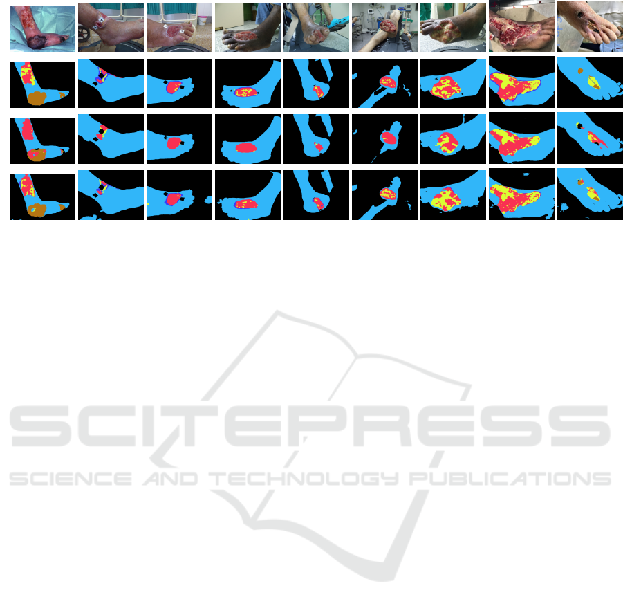
Figure 3: Results on the 7-class segmentation problem. The rows present, respectively, the input images, ground truth masks,
segmentation by the model trained without TL nor DA, and by the model with TL and DA.
The use of transfer learning and data augmentation
helped to increase the F1 and IoU scores of U-Net on
almost all classes (Table 3). Unfortunately, the ran-
dom split performed to compose the training, valida-
tion, and test subsets prevented the rarest case of tis-
sue (i.e., exposed tendon) from being identified by U-
Net, ENet, DeepLab, and SegNet. We have only three
images with an exposed tendon in our dataset, and the
small area of this tissue tends to become unimpres-
sive when the image is reduced to 224 × 224. How-
ever, this tissue is not as important in DFU analysis
as are epithelization () and granulation (), which
indicate advances in ulcers healing, or slough () and
necrotic () tissues, which represent barriers to re-
covery ulcers. By comparing the ground truth masks
(Figure 3, second row) to segmentation result, its is
clear that the , , , and regions of the DFUs
were better segmented using data augmentation com-
bined with transfer learning (fourth row) than without
the use of these techniques (third line).
4.2 Registration Results
The naive matching of descriptors computed by the
U-Net leads to a large number of inconsistent corre-
spondence pairs. For instance, note the crossed lines
in the first column of examples shown in Figure 4. In
all the cases analyzed we have noticed that the ratio
between the amount of incorrect and correct matches
changes favorably after RANSAC found the funda-
mental matrix that best fits the data. For example, in
the second column in Figure 4, the remaining sets of
matches are more consistent than the sets presented
in the first column. The number of matching pairs
dropped from 670 to 507 in the first row and 806 to
92 in the second row. Recall that sparse correspon-
dence is not sufficient to carry out image registration.
The last column of Figure 4 shows examples of dense
registration achieved by the last step of our approach.
The use of the angular weights combined with epipo-
lar constraints was key in finding coherent dense cor-
respondences because some descriptors are similar,
especially on healthy skin which is poor in texture.
The angular weighting showed good efficiency in the
elimination of outliers.
5 CONCLUSIONS
This paper presented a DFU image dataset and a seg-
mentation and registration procedure for such images.
Our dataset includes 222 images and their respective
segmentation masks considering six different types of
tissues, plus background. We first trained the segmen-
tation network assuming the wound and non-wound
classes of different public datasets and then applied
transfer learning to extend the classification to the 7-
class semantic segmentation in our dataset. The reg-
istration process is based on the comparison of visual
clues encoded by features computed by the encoder of
the U-Net used for segmentation and geometrical con-
straints from the epipolar geometry of the cameras.
The execution of both segmentation and registration
methods performed well, especially considering that
the wound of the same patient changes in appearance
during treatment, making the dense registration more
challenging. Our experiments proved that training
U-Net models with small DFU datasets is sufficient
to obtain feature descriptors representative enough to
perform DFU image registration. This is an exciting
result of our work, as other authors (e.g., Long et al.
(2014); Dara and Tumma (2018)) have pointed out
that large datasets are needed to produce representa-
tive features for natural images.
VISAPP 2022 - 17th International Conference on Computer Vision Theory and Applications
516
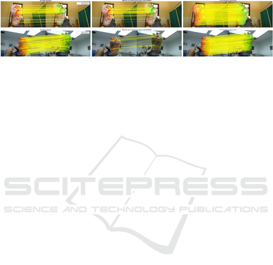
Figure 4: From the left to the right: sparse matching of features, consensus set returned by RANSAC, and dense registration.
We present 1% of the correspondences to avoid clutter.
Future work include monitoring the evolution of
wounds over time by calculating tissue area variations
from the dense registration. Also, we are working on
an attention mechanism to guide the model to classify
pixels as DFU tissue, healthy skin, or background and
then segment the five types of DFU tissues.
ACKNOWLEDGEMENTS
Y. Toledo and L. A. F. Fernandes are partially funded
by Google LARA 2020. L. A. F. Fernandes is also
funded by a FAPERJ grant (E-26/202.718/2018), and
Y. Toledo is also funded by a CAPES fellowship.
REFERENCES
Badrinarayanan, V., Kendall, A., and Cipolla, R. (2017).
SegNet: a deep convolutional encoder-decoder archi-
tecture for image segmentation. PAMI, 39(12):2481–
2495.
Bergstra, J., Yamins, D., and Cox, D. (2013). Making a
science of model search: hyperparameter optimization
in hundreds of dimensions for vision architectures. In
Proc. ICML, pages 115–123.
Chen, L. C., Papandreou, G., Schroff, F., and Adam, H.
(2017). Rethinking atrous convolution for semantic
image segmentation. arXiv:1706.05587.
Dara, S. and Tumma, P. (2018). Feature extraction by using
deep learning: a survey. In Proc. ICECA, pages 1795–
1801.
Goyal, M., Yap, M. H., Reeves, N. D., Rajbhandari, S., and
Spragg, J. (2017). Fully convolutional networks for
diabetic foot ulcer segmentation. In Proc. SMC, pages
618–623.
Hao, S., Zhou, Y., and Guo, Y. (2020). A brief survey on se-
mantic segmentation with deep learning. Neurocom-
puting, 406:302–321.
Hartley, R. and Zissermann, A. (2004). Multiple view geom-
etry in computer vision. Cambridge University Press,
2nd edition.
Kaswan, K. E., Jamil, N., and Roslan, R. (2020). Deep
learning on wound segmentation and classification: a
short review and evaluation of methods used. South-
east Eur. J. Soft Comput., 9(2):6–10.
Klein, S., Staring, M., Murphy, K., Viergever, M. A., and
Pluim, J. P. W. (2009). Elastix: a toolbox for intensity-
based medical image registration. Trans Med Imaging,
29(1):196–205.
Li, L., Jamieson, K., DeSalvo, G., Rostamizadeh, A., and
Talwalkar, A. (2017). Hyperband: a novel bandit-
based approach to hyperparameter optimization. J.
Mach. Learn. Res., 18(1):6765–6816.
Long, J. L., Zhang, N., and Darrell, T. (2014). Do convnets
learn correspondence? In Proc. NeurIPS, pages 1601–
1609.
Medetec (2021). Medetec wound database. [Online].
Available: https://medetec.co.uk/slide\%20scans/
foot-ulcers/index.html.
Paszke, A., Chaurasia, A., Kim, S., and Culurciello, E.
(2016). ENet: a deep neural network architecture for
real-time semantic segmentation. arXiv:1606.02147.
Ronneberger, O., Fischer, P., and Brox, T. (2015). U-Net:
convolutional networks for biomedical image segmen-
tation. In Proc. MICCAI, volume 9351, pages 234–
241.
Sol
´
ıs-S
´
anchez, L. O., Ortiz-Rodriguez, J. M., Casta
˜
neda-
Miranda, R., Martinez-Blanco, M. R., Ornelas-
Vargas, G., Galv
´
an-Tejada, J. I., Galv
´
an-Tejada, C. E.,
Celaya-Padilla, J. M., and Casta
˜
neda-Miranda, C. L.
(2016). Identification and evaluation on diabetic foot
injury by computer vision. In Proc. ICIT, pages 758–
762.
Wagh, A., Jain, S., Mukherjee, A., Agu, E., Pedersen, P. C.,
Strong, D., Tulu, B., Lindsay, C., and Liu, Z. (2020).
Semantic segmentation of smartphone wound images:
comparative analysis of AHRF and CNN-based ap-
proaches. IEEE Access, 8:181590–181604.
Wang, C., Anisuzzaman, D. M., Williamson, V., Dhar,
M. K., Rostami, B., Niezgoda, J., Gopalakrishnan, S.,
and Yu, Z. (2020). Fully automatic wound segmenta-
tion with deep convolutional neural networks. Scien-
tific Reports, 10(1):1–9.
Wang, C., Anisuzzaman, D. M., Williamson, V., Dhar,
M. K., Rostami, B., Niezgoda, J., Gopalakrish-
nan, S., and Yu, Z. (2021). FUSeg dataset.
[Online]. Available: https://github.com/uwm-bigdata/
wound-segmentation/.
Zhang, X., Gilliam, C., and Blu, T. (2019). Parametric
registration for mobile phone images. In Proc. ICIP,
pages 1312–1316.
U-Net-based DFU Tissue Segmentation and Registration on Uncontrolled Dermoscopic Images
517
