
Innovative Methodology for the 3D Reconstruction of Body
Geometries using Open-source Software
Javier Tuesta-Guzmán
1a
, William Solórzano-Requejo
1,2 b
, Gustavo Grosso-Salazar
1c
,
Carlos Ojeda
1d
and Andrés Díaz Lantada
2e
1
Department of Mechanical and Electrical Engineering, Universidad de Piura, Piura, Peru
2
ETSI Industriales, Universidad Politécnica de Madrid, Madrid, Spain
carlos.ojeda@udep.edu.pe, andres.diaz@upm.es
Keywords: 3D Scan, Reverse Engineering, Meshroom, Open-source Software, Photogrammetry.
Abstract: Bioengineering teaching has limitations in developing countries due to the inaccessibility of expensive
technology like scanners and commercial software, which holds back progress in the biomedical area because
of a lack of resources. In this work, a new methodology is presented with the aim of obtaining a 3D model of
body part by open-source software: Meshroom
®
, Meshmixer
®
, Ultimaker Cura
®
; and a cell phone camera.
The procedure is based on three methods which were tested: images taken for a short time, burst mode and
video-to-frames. Through the process of reverse engineering photogrammetry, an arm and a foot were
obtained from images and for comparing the model with the real body part, 3D printing was used. The
outstanding method is video-to-frames thanks to the high quality of the generated models and the shortest
reconstruction time it presents. The technique developed can promote the education of engineers in the
biomedical area, also providing an advance for developers with low economic resources, allowing them to
have a new possibility of research.
1 INTRODUCTION
Medical Reverse Engineering (MRE) is highly used
to treat fracture problems, obtaining hurt body parts
in 3D format as STL (Stereo Lithography) or OBJ
(Object) file (Bhatti et al., 2018). MRE is also
fundamental in any medical device personalization
strategy (Ahluwalia et al., 2022). Computer-aided
design software tend to be expensive, which may
limit equal accessibility to personalized healthcare
technologies. Fortunately, different open-source
hardware and software solutions are being developed,
which synergize with the "maker's movement" and
promote equity in all fields of product development.
These technologies allow getting the affected zone,
necessary for the design and manufacture of custom
medical devices. The usage of open-source software
is a proposal for the education framework of
a
https://orcid.org/0000-0002-6923-7263
b
https://orcid.org/0000-0002-2989-9166
c
https://orcid.org/0000-0002-7570-5609
d
https://orcid.org/0000-0001-6163-5382
e
https://orcid.org/0000-0002-0358-9186
engineering students due to remote education as a
consequence of the COVID 19 pandemic, which
forced to change the techniques used to teach
(Pokhrel & Chhetri, 2021). Any researchers already
focused their attention on the development of
technologies that could assist students in anatomy
remote education (Iwanaga et al., 2021) as Qlone
®
for
3D scanning.
Scanning in the biomedical area is a method to
obtain an external body part like legs, arms, hands,
face; necessary to design an external fixation for
closed fracture (Alqahtani et al., 2021) or help relieve
articular injuries (Munoz-Guijosa et al., 2020) but for
this, designers need a special camera to scan it, and
this is a limitation for the developed of personalized
medical technology due to the high cost (Le et al.,
2010). Fortunately, Meshroom
®
(Griwodz et al.,
2021) and COLMAP
®
(Schonberger & Frahm, 2016)
162
Tuesta-Guzmán, J., Solórzano-Requejo, W., Grosso-Salazar, G., Ojeda, C. and Díaz Lantada, A.
Innovative Methodology for the 3D Reconstruction of Body Geometries using Open-source Software.
DOI: 10.5220/0010870200003123
In Proceedings of the 15th International Joint Conference on Biomedical Engineering Systems and Technologies (BIOSTEC 2022) - Volume 1: BIODEVICES, pages 162-169
ISBN: 978-989-758-552-4; ISSN: 2184-4305
Copyright
c
2022 by SCITEPRESS – Science and Technology Publications, Lda. All rights reserved
exist, whose principal function is to do the object
reconstruction from images. This technique is called
photogrammetry and is less common for 3D
reconstruction but a MRE useful tool to get the base
for the biodevices design (Struck et al., 2019). The
photogrammetry for 3D surface scanning allows good
visualization of the objects and people without a high
knowledge about photography can do a
reconstruction (Grabherr et al., 2016).
This project explores innovative approaches to
promote MRE through open-source software. A
procedure relying on the combination of varied
software, including: VLC Media Player 3.0.16
Vetinari
®
, Meshroom 2021.1.0
®
, Meshmixer
3.5.474
®
, and UltimakerCura 4.8.0
®
, is presented. To
the author’s knowledge, this combination is
innovative and reported for the first time. It is
illustrated through a set of case studies.
The goal is to promote collaborative work,
inspired in projects as UBORA (De Maria et al.,
2020), wanting to reinforce the techniques used for
remote study and have an important impact in
developing countries for people with few resources,
focused on the formation of students, engineers and
doctors who want to research in the biomedical field.
This work is organized as follows: first, materials
and methods used to obtain the arm and foot models
will be explained; secondly, the results and discussion
of the quality of models, and finally the conclusions
of this project.
2 MATERIALS AND METHODS
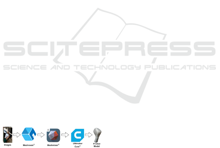
Figure 1 shows the roadmap to obtain the 3D model,
as a base for designing personalized medical devices,
from images of the body part employing open-source
software. This research assesses the influence of the
method to get the images, in the mesh given by the
programs.
Figure 1: Roadmap.
External devices need a geometrical body
obtained through photogrammetry, which needs
images around the body part, then they are imported
to Meshroom
®
which reconstructs the scene and
generates an OBJ file, different tests will be done to
define which is the best method together with the
correct number of images. Subsequently,
Meshmixer
®
was used to optimize, crop, and repair
the 3D model that finally is prepared and printed
through Ultimaker Cura
®
.
2.1 Reverse Engineering
2.1.1 Photogrammetry
Photogrammetry can be understood as a methodology
to determine distance relations between different
bodies in space from images. One image provides
two-dimensional coordinates of each point of the
photo, and if it is complemented by a second image
from a different position, so a three-dimensional
coordinate could be calculated for each point (Linder,
2009). It is a powerful tool for reverse engineering to
obtain models of inanimate objects, also could be
used to reconstruct body parts with the correct steps.
Meshroom
®
is an open-source software for 3D
reconstructions based on the AliceVision framework.
The software works in a base of photogrammetry, and
to make a reconstruction is necessary to configure
nodes that Meshroom
®
offers. The environment
nodes by default receive the inputs (images),
separating them into groups and highlighting the most
notable features, then generate a points cloud of the
scene in 3D, filter the inconsistencies and finally
generate a mesh and project the textures in the model
(Dong et al., 2021). Meshroom
®
will be used for the
experiments of bodies part reconstruction adding a
new node to establish the actual scale.
Their minimum requirements are RAM: 8GB,
GPU: NVIDIA CUDA, CPU: Intel or AMD and
works in any operating system.
To take the pictures, it is necessary to use a camera
or multiple cameras (Peyer et al., 2015) which allows
obtaining a detailed view of the body reconstructed.
The cell phone used was a Samsung A50 which has a
triple camera. The primary camera has a resolution of
24.9MP with an aperture of f/1.7. The second (ultra-
wide) and third (depth sensor) ones have an aperture
of f/2.0 with 8MP and 5MP respectively.
2.1.2 Meshroom and Animated Objects
Through Meshroom
®
, different models can be
obtained of all types of inanimate things as vehicles
(Matys et al., 2021), ship structures (Shah et al.,
2021), and more if given it images of the object from
360° degrees as shown in Figure 2. Meshroom
®
uses
these images to establish measure relations between
the background and the main piece. For this reason,
while more images are given, the detail of the final
model should be better, but not all is quantity because
Innovative Methodology for the 3D Reconstruction of Body Geometries using Open-source Software
163
if blurred images are used, the final model will be
affected. The difficulty is obtaining a 3D high-quality
model of a self-moving object.
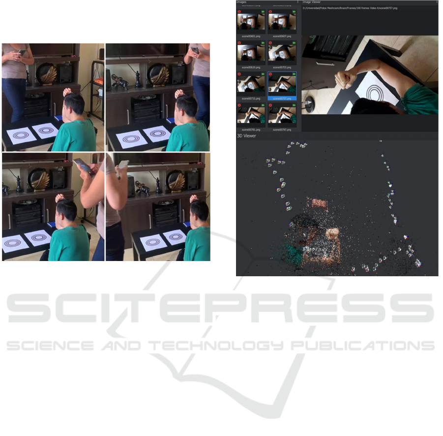
Figure 2: Images surrounds the body.
For example, arm, face, foot, hand, and functional
body parts are considered as self-moving objects, if
someone tries to take images surrounding them, it is
a fact that exists minimal involuntary movements
because of the tired of maintaining these parts in a
special position, necessary to do the images for 2
minutes or more. If these images are given to
Meshroom
®
, the final model will have all the
background: walls, floor, and other objects minus the
body part. An idea could be to eliminate the
background and only have the body part in the
pictures; the result will be an amorphous model
because distance relations are eliminated, which are
essential for Meshroom
®
works. Therefore, it is
necessary that the interaction with the participants be
the minimum to avoid these unwanted movements
(Peyer et al., 2015).
Figure 3 shows the Meshroom
®
interface; the
images generate a point cloud that is exported in OBJ
format. The default model scale is not the real
magnitude. For this reason, it is necessary to use the
CCTAG3 templates (CCTag /MarkersToPrint at
Develop · Alicevision/CCTag, 2021) that were
provided by AliceVision, since Meshroom
®
needs to
set up the real size.
Figure 3: Meshroom
®
interface.
2.2 Experiments
Six experiments have been performed with different
methods and several images to generate the best
model as possible. The first method consisted in
taking pictures surrounding the body; the
reconstructions were made with 100 images taken for
4 minutes approximately, then, this experiment was
repeated with half of the images (50) that were taken
from the first 2 minutes. As some involuntary
movements may exist, another approach to take the
pictures should be sought, for this purpose, in the
second method the use of the burst mode of the phone
was performed. Finally, the third method (video-to-
frames), proposed by the authors, is based on
recording a 40-second video, since Meshroom
®
does
not use videos for reconstruction it must be converted
into frames. To maintain the conditions of each
reconstruction, 100 and 50 images were used for each
method.
To convert the video on frames, VLC Media
Player
®
was used (Figure 4). This program generates
a high number of images according to the recording
ratio, for example, 100 frames in 30-second video.
Thus, the number of images for the recognition is
maximized and the involuntary movements are
minimized.
BIODEVICES 2022 - 15th International Conference on Biomedical Electronics and Devices
164
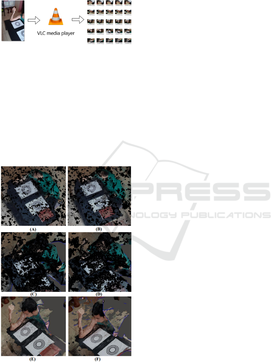
Figure 4: Conversion of video-to-frames through VLC
Media Player
®
.
3 RESULTS AND DISCUSSION
3.1 Arm
To take the images, the person was sitting down with
their arm supported on a table where the CCTAG3
templates stay collocated with 30cm of distance and
should be visible.
The models were exported from Meshroom® and
imported in Meshmixer® to visualize it, for
understanding the behavior of the models, a similar
sight of the reconstruction will be compared.
Figure 5: Arm reconstruction with (A) 100 and (B) 50
photos, (C) 100 and (D) 50 burst photos, (E) 100 and (F) 50
frames.
Figure 5 shows the different models obtained
through the experiments. In Figure 5A is visible that
the arm was not reconstructed but at least obtain a part
of the neck, shoulder, and a few of the inanimate
objects. In Figure 5B, despite using half of the photos
on purpose to reduce the time to take images (4 to 2
minutes), the result was not good, this model does not
show the arm too. In Figure 5C appears a form similar
to the arm with poorly defined parts (errors in
reconstruction), but the shape is noticeable. In Figure
5D, the use of half of the images did a few differences
defining better some parts of the body but not enough.
Figure 5E presents the arm reconstructed with a void
in the last part of the forearm and Figure 5F present
the arm with nom-uniform areas. Anyway, the
difference with the first four models is noticeable.
In the models from method of photos is notorious
that the presence of the involuntary movements is the
principal problem, for this reason, images should not
be taken for a long time or else not be obtained the
desired model, despite this, it is a necessity a large
number of images for the reconstruction. Burst mode
of phone method presents better models than only
taking photos due to reduction of the capture time, but
not enough to minimize the effect of the involuntary
movements. Also, any images from burst mode could
have low quality when trying surround the main piece
and as a consequence the final models are poorly
detailed and useless. For video-to-frames method was
obtained a good reconstruction, first, the model is
recognizable, there is no doubt that the reconstruction
is an arm; second, the arm was reconstructed
completely and with high quality compared with the
previous methods, third, the surroundings objects
were reconstructed correctly too. It is visible that
these models present non-uniform areas due to in
frames exist a problem of brightness, the recording
place has angles where the arm looks darker, and
deformations occur right at these angles. In other
words, when existing different illumination, the
model will be affected directly, so the lightning must
be constant to preserve the quality of reconstruction.
It visualized that the use of half images has fewer
errors in the result obtained in each technique, for
example, 50 bursts model is better than 100 bursts
model; this may be due to if use an excessive number
of images, results in oversizing whose inconsistencies
are not suppressed in totally in filter nodes.
Table 1 summarizes the results for each
experiment through Meshroom
®
. It is easily noticed
that the best model was obtained with the method
proposed “video-to-frames”, also the number of
triangles in Table 1 has a relation with the number of
Innovative Methodology for the 3D Reconstruction of Body Geometries using Open-source Software
165
images used, more photos make more reconstruction
of the main piece and its environment.
Table 1: Vertices, triangles, and reconstruction time for
each arm model.
Model Vertices Triangles Time
100 photos 308672 612916 60m 7s
100 bursts 428034 849489 86m 50s
100 frames 138060 274218 36m 56s
50 photos 232459 461799 28m 8s
50 bursts 366186 727762 45m 57s
50 frames 37695 74290 10m 11s
Models with 50 pictures have fewer triangles than
the model with their respective method with 100. It is
not possible to compare the number of triangles of the
six models only having the arm, cropping the OBJ
file, because in some the arm is not completely
reconstructed.
The fastest reconstruction for 50 and 100 images
was the video-to-frames models, so this method
performs a better reconstruction in less time.
3.2 Foot
The experiments were repeated with the foot to
analyze the performance with another body region,
for comparative
purposes. In this case, to took
images, the person stood up and placed their leg near
to CCTAG3 templates which establishes the real
scale.
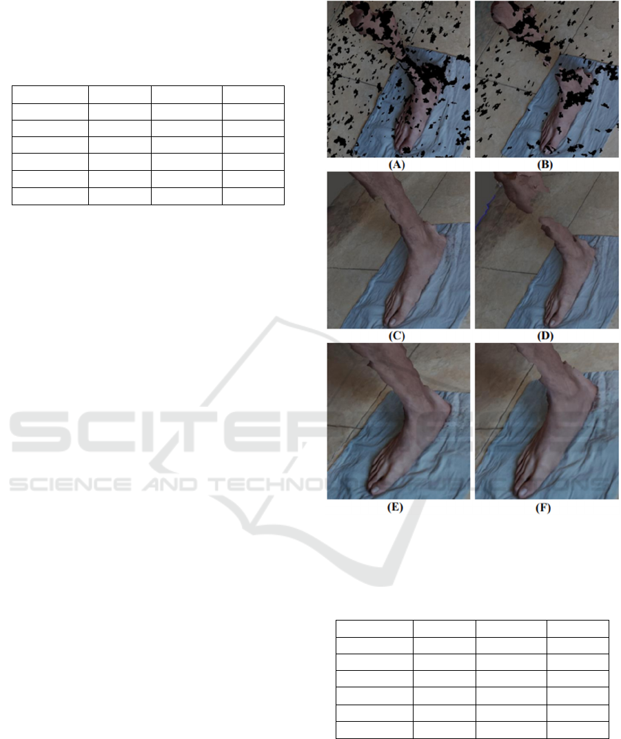
Figure 6 shows the results of the reconstruction of
each technique and the respective number of images.
Figure 6A presents an amorphous and incomplete
mesh, same problem in Figure 6B but adding that due
to the fewer images, the reconstruction presents a gap
between the foot and calf. For Figure 6C, the calf zone
is visible with protuberances, and it is the same for
the model of Figure 6D but with a gap in the leg. In
Figure 6E, the reconstruction is similar to the 100
burst model but more complete and better defined.
Finally, Figure 6F presents a good quality model but
with more protuberances in the calf zone. Same as the
models of the arm, the best model was generated by
the video-to-frames method.
The results are shown in Table 2. The model with
the fewest triangles and the best reconstruction time
was from video-to-frames. The number of triangles
has a direct impact on the reconstruction time. As this
is minimal, less computational resources are required,
which allows the use of this technique and technology
for educational purposes.
Figure 6: Foot reconstruction with (A) 100 and (B) 50
photos, (C) 100 and (D) 50 burst photos, (E) 100 and (F) 50
frames.
Table 2: Vertices, triangles, and reconstruction time for
each foot model.
Model Vertices Triangles Time
100 photos 477171 949084 94m 16s
100 bursts 461408 918505 76m 6s
100 frames 190621 379804 38m 36s
50 photos 406215 808415 51m 56s
50 bursts 417762 816884 58m 58s
50 frames 138283 275557 15m 43s
Best models will be selected for arm and foot on
purpose to be optimized with Meshmixer
®
and printed
with Ultimaker Cura
®
. Both are the models obtained
with 50 frames due to being better reconstruction,
with minor time and weight file.
Meshroom
®
delivers unsmoothed models with
some surface defects, for this is necessary to use CAD
BIODEVICES 2022 - 15th International Conference on Biomedical Electronics and Devices
166
software to eliminate this problem. Meshmixer
®
is an
open-source software useful to optimize OBJ/STL
files and will be used to visualize and fix our models.
To optimize arm and foot, both were cropped and
just stayed with body part of interest (Figure 7). Auto-
repair Meshmixer
®
tools were used to solve mesh
problems if they exist. After, the models are exported
as STL files.
Figure 7: Cropping and optimization with Meshmixer
®
for
the (A) arm and (B) foot models.
To obtain the physical model, the 3D printing
technique used was Fused Material Deposition
(FDM). Between the steps of this process was
included the lamination in several layers of the STL
models using the Ultimaker Cura
®
slicer. The G Code
was exported from Ultimaker
®
to manufacture the 3D
models employing Ender 3 Pro
®
printer.
The parameters used for the impression were the
following: PLA Flibox Silver, infill 10% and infill
pattern line, wall line count 3, bottom and top layers
4, speed 50 mm/s, retraction activate, fan speed
100%. Supports were used for the foot model.
Figure 8: Comparison between the printed and real arm.
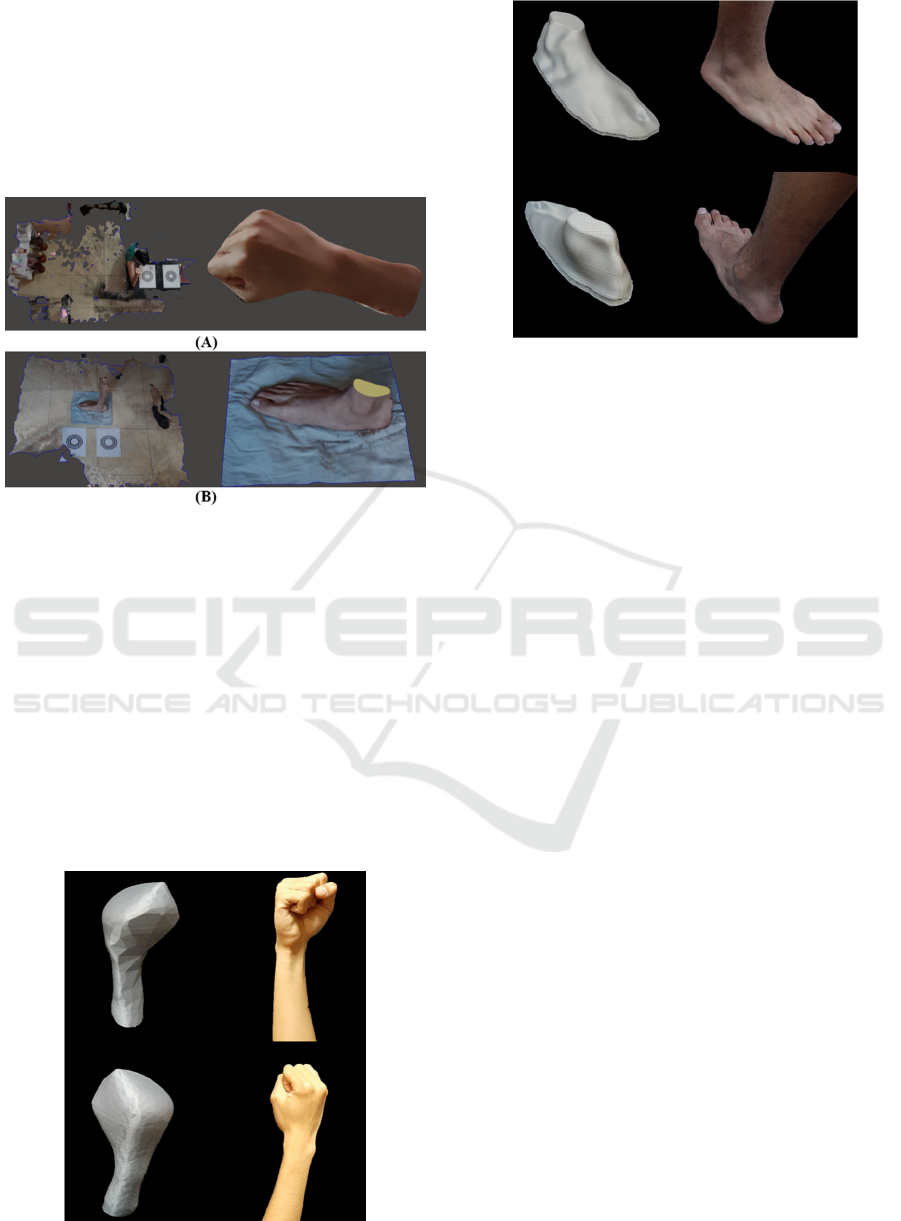
Figure 9: Comparison between the printed and real foot.
Figures 8 and 9 show two views of the 3D printed
model compared with the scanned body for arm and
foot respectively. Printed models are similar to real
animated objects in shape and scale, the printed arm
has the geometry of the forearm, and the fingers are
unified due to Meshroom recognizing them as one
body. For printed foot, the geometry matches with the
real foot. Both models have good quality and if this
methodology continues to be refined, it can be used
to obtain the base bodies for the design of 3D printing
orthoses (Górski et al., 2020), orthopaedic insoles
(Cendrero et al., 2021; Shalamberidze et al., 2021)
and facial protection masks (Morita et al., 2007).
Other authors (Peyer et al., 2015) constructed a
system with 18 cameras to perform 3D reconstruction
from taking simultaneous photos, minimizing the
interaction time with the body. Their surface mesh
presents high-resolution, but it is not easy for students
from developing countries to obtain these systems.
The images used for these experiments had a
quality of 25 MP and the results were good, if the
number of pixels was increased, the results would
have more detail and a better surface finish.
4 CONCLUSIONS
Printed models of OBJ/STL files generated with the
proposed methodology have high quality for
educational purposes and transmit the essence of the
main piece coinciding with reality. This opens a
world of possibilities for the reconstruction of
animated objects from images as the design of
biodevices or remote education through laboratories
in study centres for the reconstruction of 3D models
without an expensive camera.
Innovative Methodology for the 3D Reconstruction of Body Geometries using Open-source Software
167

The video-to-frames technique proved more
reliable than only photos and burst mode. Their
models show the essence of the shapes in both prints,
also minimize the time for the reconstruction but still
have a few protuberances due to the change of
illumination concerning the angle of the shot and the
limited number of pixels of the images. The method
still could be better if images are taken in other
prepared places where the lighting remains constant
facilitating the reconstruction process and if a
professional camera will be employed to record the
video. Between the data used, exists blurry images
which could be eliminated manually, but exist
algorithms that can do this automatically like Fast
Fourier Transform or Variance of Laplacian which
can help to improve the proposed technique in the
future.
Among main limitations of the study, it is
important to mention that a simple camera has been
used, which may affect precision, although at the
same time its use puts forward the possibility of using
very low-cost hardware and software for promoting
MRE. Regarding future studies, our proposal is to
progress in processes for automated generation of 3D
models, to enhance precision and to employ these and
similar reconstructions, as input for the design of
personalized medical devices, such as splints, insoles,
braces and multiple orthoses.
ACKNOWLEDGEMENTS
The authors would like to thank the support of the
Biomechanics Group of the Universidad de Piura and
to the Product Development Laboratory of the
Universidad Politécnica de Madrid. Also, we
acknowledge the support of reviewers and their
relevant recommendations, which help to do a more
consistent and detailed paper.
REFERENCES
Ahluwalia, A., De Maria, C., & Díaz Lantada, A. (Eds.).
(2022). Engineering Open-Source Medical Devices (1st
ed.). Springer International Publishing.
https://link.springer.com/book/9783030793623
Alqahtani, M. S., Al-Tamimi, A. A., Hassan, M. H., Liu, F.,
& Bartolo, P. (2021). Optimization of a Patient-Specific
External Fixation Device for Lower Limb Injuries.
Polymers, 13(16), 2661. https://doi.org/10.3390/
polym13162661
Bhatti, A., Syed, N. A., & John, P. (2018). Chapter 5—
Reverse Engineering and Its Applications. In D. Barh
& V. Azevedo (Eds.), Omics Technologies and Bio-
Engineering (pp. 95–110). Academic Press.
https://doi.org/10.1016/B978-0-12-804659-3.00005-1
CCTag/markersToPrint at develop · alicevision/CCTag.
(2021). GitHub. https://github.com/alicevision/CCTag
Cendrero, A. M., Fortunato, G. M., Munoz-Guijosa, J. M.,
De Maria, C., & Díaz Lantada, A. (2021). Benefits of
Non-Planar Printing Strategies Towards Eco-Efficient
3D Printing. Sustainability, 13(4), 1599.
https://doi.org/10.3390/su13041599
De Maria, C., Di Pietro, L., Lantada, A. D., Ravizza, A.,
Mridha, M., Torop, J., Madete, J., Makobore, P., &
Ahluwalia, A. (2020). The UBORA E-Infrastructure for
Open Source Innovation in Medical Technology. In J.
Henriques, N. Neves, & P. de Carvalho (Eds.), XV
Mediterranean Conference on Medical and Biological
Engineering and Computing – MEDICON 2019 (pp.
878–882). Springer International Publishing.
https://doi.org/10.1007/978-3-030-31635-8_106
Dong, C., Liang, W., & Xu, P. (2021). 3D refined
reconstruction for brushes based on multiple images.
2021 3rd International Conference on Advances in
Computer Technology, Information Science and
Communication (CTISC), 309–314. https://doi.org/
10.1109/CTISC52352.2021.00063
Górski, F., Wichniarek, R., Kuczko, W., Żukowska, M.,
Lulkiewicz, M., & Zawadzki, P. (2020). Experimental
Studies on 3D Printing of Automatically Designed
Customized Wrist-Hand Orthoses. Materials, 13(18),
4091. https://doi.org/10.3390/ma13184091
Grabherr, S., Baumann, P., Minoiu, C., Fahrni, S., &
Mangin, P. (2016). Post-mortem imaging in forensic
investigations: Current utility, limitations, and ongoing
developments. Research and Reports in Forensic
Medical Science, 6, 25–37. https://doi.org/10.2147/
RRFMS.S93974
Griwodz, C., Gasparini, S., Calvet, L., Gurdjos, P., Castan,
F., Maujean, B., De Lillo, G., & Lanthony, Y. (2021).
AliceVision Meshroom: An open-source 3D
reconstruction pipeline. Proceedings of the 12th ACM
Multimedia Systems Conference, 241–247.
https://doi.org/10.1145/3458305.3478443
Iwanaga, J., Terada, S., Kim, H.-J., Tabira, Y., Arakawa,
T., Watanabe, K., Dumont, A. S., & Tubbs, R. S.
(2021). Easy three-dimensional scanning technology
for anatomy education using a free cellphone app.
Clinical Anatomy, 34(6), 910–918. https://doi.org/
10.1002/ca.23753
Le, C., Jos, V. S., Le, T. H., Lam, K., Soe, S., Zlatov, N.,
Le, T. P., & Pham, D. T. (2010). Medical reverse
engineering applications and methods [Conference
Proceedings]. University of Greenwich; INCDMTM.
https://gala.gre.ac.uk/id/eprint/11735/
Linder, W. (2009). Introduction. In W. Linder (Ed.), Digital
Photogrammetry: A Practical Course (pp. 1–17).
Springer. https://doi.org/10.1007/978-3-540-92725-9_1
Matys, M., Krajcovic, M., & Gabajova, G. (2021). Creating
3D models of transportation vehicles using
photogrammetry. Transportation Research Procedia,
55, 584–591. https://doi.org/10.1016/j.trpro.2021.07.0
25
BIODEVICES 2022 - 15th International Conference on Biomedical Electronics and Devices
168

Morita, R., Shimada, K., & Kawakami, S. (2007). Facial
Protection Masks After Fracture Treatment of the Nasal
Bone to Prevent Re-injury in Contact Sports. Journal of
Craniofacial Surgery, 18(1), 143–145. https://doi.org/
10.1097/01.scs.0000246729.23483.87
Munoz-Guijosa, J. M., Zapata Martínez, R., Martínez
Cendrero, A., & Díaz Lantada, A. (2020). Rapid
Prototyping of Personalized Articular Orthoses by
Lamination of Composite Fibers upon 3D-Printed
Molds. Materials, 13(4), 939. https://doi.org/
10.3390/ma13040939
Peyer, K. E., Morris, M., & Sellers, W. I. (2015). Subject-
specific body segment parameter estimation using 3D
photogrammetry with multiple cameras. PeerJ, 3, e831.
https://doi.org/10.7717/peerj.831
Pokhrel, S., & Chhetri, R. (2021). A Literature Review on
Impact of COVID-19 Pandemic on Teaching and
Learning. Higher Education for the Future, 8(1), 133–
141. https://doi.org/10.1177/2347631120983481
Schonberger, J. L., & Frahm, J.-M. (2016). Structure-from-
Motion Revisited. 2016 IEEE Conference on Computer
Vision and Pattern Recognition (CVPR), 4104–4113.
https://doi.org/10.1109/CVPR.2016.445
Shah, F. M., Gaggero, T., Gaiotti, M., & Rizzo, C. M.
(2021). Condition assessment of ship structure using
robot assisted 3D-reconstruction. Ship Technology
Research, 0(0), 1–18. https://doi.org/10.1080/09377
255.2021.1872219
Shalamberidze, M., Sokhadze, Z., & Tatvidze, M. (2021).
The Design of Individual Orthopedic Insoles for the
Patients with Diabetic Foot Using Integral Curves to
Describe the Plantar Over-Pressure Areas.
Computational and Mathematical Methods in
Medicine, 2021, e9061241. https://doi.org/10.1155/
2021/9061241
Struck, R., Cordoni, S., Aliotta, S., Pérez-Pachón, L., &
Gröning, F. (2019). Application of Photogrammetry in
Biomedical Science. In P. M. Rea (Ed.), Biomedical
Visualisation: Volume 1 (pp. 121–130). Springer
International Publishing. https://doi.org/10.1007/978-
3-030-06070-1_10
Innovative Methodology for the 3D Reconstruction of Body Geometries using Open-source Software
169
