
The Potential Application in Medical Biomimetic Materials
Wangcheng Zhu
a
Shanghai Pinghe, Bilingual School, Shanghai, China
Keywords: Biomimetic Material, Spider Silk, Nacre, Bone, Nanomaterials.
Abstract: The "biomimetic materials science" formed by the intersection of life science and material science has
excellent theoretical and practical significance. Biomimetic materials science takes material formation and
structure as the target, considers artificial material at the view of biomaterial, exploring the manufacture and
design of material from the angle of biological function. With unique formation methods, structural
characteristics, and biomechanical properties, biomimetic materials are becoming a new field of materials
science. Based on a lot of research work, this article reviews and summarizes the work done by predecessors
in medical bionic materials. The review focuses on the research progress of biomimetic materials science
includes silk protein, nacre, bone, nanomaterials. Numerous studies have shown that these biomimetic
materials have a potential application in medicine. Finally, the development prospects of medical
biomimetic materials are forecasted.
1 INTRODUCTION
1
In recent years, with the development of related
disciplines and the progress of modern technology,
bionic materials have been developed rapidly. The
results were widely used in aviation materials,
biomedical materials, textile materials and so on.
Biomimetic materials science aims to clarify
biological materials' structure and formation process,
with biological materials to consider the idea of
artificial materials, from the perspective of
biological function to consider the material design
and production.
Biomimetic material science is an essential
branch of bionics, which refers to study the
structural characteristics, structure, and activity of
biological materials from the molecular level. It
aims to find the new science similar or better than
the original biological materials, chemistry, material
science, biology, physics, and other disciplines
cross. Medical bionic materials are an important
application field of bionic materials.
Bionic materials, such as artificial tooth enamel,
artificial bone materials, and bionic artificial fibers,
through the design of structures and materials, are
expected to achieve high efficiency, low energy
consumption, and environmental harmony
a
https://orcid.org/0000-0002-7040-353X
(Arcidiacono, 2002). Imitation of natural animal and
plant-specific function and intelligent response, the
development of and biological similarity or beyond
the existing biological function of artificial
materials, such as lotus leaf self-cleaning materials,
like the shark's self-lubricating material, in the gene
transformation of the cells in the efficient synthesis
of chiral molecules and macromolecules (Tabata,
2001). Imitate the structure and performance of
biological tissues and organs, study the structure and
properties of natural biological materials from the
perspective of materials science, and develop bionic
materials. This review will shed light on the research
and development of high-performance biomedical
materials. This review mainly introduces the
biomimetic materials of silk protein, bone, nacre,
and cells, summarizes the current development of
biomimetic materials, and hopes to inspire other
researchers.
780
Zhu, W.
The Potential Application in Medical Biomimetic Materials.
DOI: 10.5220/0011295100003443
In Proceedings of the 4th International Conference on Biomedical Engineering and Bioinformatics (ICBEB 2022), pages 780-784
ISBN: 978-989-758-595-1
Copyright
c
2022 by SCITEPRESS – Science and Technology Publications, Lda. All rights reserved

2 MECHANISM AND
APPLICATION OF
BIOMIMETIC MATERIALS
2.1 Biomimetic Materials of Silk
Protein
Silk protein can be used to prepare excellent
biomaterials due to its good biocompatibility, low
immunogenicity, processability, and degradability.
In recent years, based on the basic principles of
tissue engineering, through material processing
technology, various forms of silk protein scaffolds
(including three-dimensional porous scaffolds,
nanofibers, hydrogels, two-dimensional membranes,
etc.) (Applegate, 2016) and silk protein drug carriers
(Microspheres, nanoparticles, etc.) have been used in
the repair of hard bones, ligaments, skin,
cardiovascular, cartilage, cornea, nerves and other
organs and tissues and anti-cancer therapy (Melke,
2016).
According to different technologies for obtaining
silk protein, the source of silk protein can be divided
into natural silk protein, recombinant silk protein,
and genetically modified silk protein. At present,
natural silks that are used and studied more include
spider silk protein and fibroin. In spider silk protein,
major ampullate silk has the advantages of high
strength, good tensile strength, and large breaking
energy, so it is currently the most studied kind of
spider silk (Hinman, 2000). The silk fibroin in the
silkworm silk fiber is coated inside the sericin, with
a diameter of about 10 μm. Studies have found that
the secondary structure of silk protein includes four
types: β-sheet, random coil, α-helix, and β-turn
(Gray, 2016).
Silk protein biomaterials prepared by different
processing technologies have different application
forms in the field of biomedicine. Silk fibers were
originally used as surgical sutures due to their
biodegradability and good biocompatibility.
Effective weaving of silk fibers can produce
"artificial ligaments" that can be used to repair or
replace broken ligaments (Altman, 2002). XU et al.
combined silk fibroin and chitosan to prepare a
composite membrane and obtained an excellent
healing effect after covering the membrane on the
wounded part of the rat (Xu,2015). YODMUANG et
al. found that silk protein gel can effectively
promote the growth of chondrocytes and the
secretion of related collagen. The formation of the
internal network of silk protein gel is conducive to
the loading of drugs or foreign genes (Yodmuang,
2015). In addition, the most studied silk protein
scaffold is used for bone tissue repair. A study
showed that adding growth factors to the scaffold
can better repair defective tissues (Meinel,2005).
Recently, Shi et al. 3D printed silk protein and
gelatin to obtain a composite scaffold that can repair
damaged cartilage in rabbit knee joints (Shi, 2017).
2.2 Biomimetic Materials of Nacre
In order to protect molluscs from water-carried
debris and predators, the shells of organisms like
abalone have evolved into a stiff and impact-
resistant material named shell. Shells are composite
materials made by mollusks that combine inorganic
minerals (CaCO3) in the surrounding environment
with organic matter generated by themselves under
ambient temperature and pressure. The formation
process of shells is a biological mineralization
process.
The shell structure is divided into 3 layers, from
the outside to inside are stratum corneum, prismatic
layer, and nacre layer. The organic layer and the
mineral layer in the nacre are arranged alternately
under appropriate magnification. The so-called
"brick and mortar" structure can be easily observed.
Its comprehensive mechanical properties, especially
fracture toughness can be rasied by 2 to 3 orders of
magnitude than that of single-phase calcium
carbonate ceramics. The alternate laminated
arrangement of nacre aragonite crystals and an
organic matrix is the key to its high toughness.
Based on this principle, material scientists have
developed the development of imitation pearl
laminated composite materials. Based on this
principle of biomineralization, Yoo et al. prepared a
boron nitride nanosheet (BNNS)/gelatin
nanocomposite. In this study, hyperbranched
polyglycerol was used to functionalize BNNS to
enhance the bonding strength between entities. By
changing the composition and arrangement degree
of BNNSs and gelatin in the nanocomposite, the
mechanical properties of the nanocomposite can be
controlled. This adjustment of mechanical properties
can produce a material with properties similar to
human cortical bone. In vitro cell experiments show
that this artificial nacre can support the adhesion and
proliferation of adipose-derived stem cells,
indicating that it can be used in biomedicine (Yoo,
2018).
Fabrication techniques such as hot-press assisted
slip casting, freeze casting, extrusion and roll
compaction, paper-making method, and layer-by-
layer self-assembly have successfully yielded
The Potential Application in Medical Biomimetic Materials
781

materials that mimic the mechanical properties of
nacre. Yongli Zhang made SiC/AL toughening
composites with AL as soft phase and SiC as the
ceramic base (Heuer,1992). And his fracture
toughness is 2 - 5 times compared to the original
one. Yang hui made A1203 toughening composites
with carbon fiber as soft phase and A1202 as a
ceramic base, increasing its fracture toughness by
1.5 -2 times (Qian,2004).
2.3 Biomimetic Materials of Bone
The mimicking of the porous and branched structure
of bone is general in the work of some architects,
originating structures that are tough and lightweight.
Bone has evolved to protect their vital organs and
provide efficient structural support to vertebrates.
Bone structure is ideal for optimal solid structure
(Almqvist, 1999)—the shape of the long bones of
the animals at both ends of the thick, elongated
intermediate. The dumbbell head in the ends of the
long bone can increase the tensile strength and
fracture toughness. Inspired by this, people design
short fiber into a "dumbbell", improving the
composite strength and elongation. It is conducive to
the coordinated movement of the material in the
fiber and the bonding materials, greatly improving
the service life.
Hu et al. developed the absorbable
chitosan/hydroxyapatite composite bionic bone
structure of fracture fixation material by situ
precipitation method and shaping not only is a
dumbbell-shaped structure but easy degradation and
absorption, with releasing orthophosphate and
calcium ion. At the same time, mechanical
properties of the biomimetic composites, such as
bending strength, bending modulus, shear strength,
compression strength, are 2 to 3 times higher than
the natural bone. It is expected to replace the metal
and become the internal fixation of bone fracture,
avoiding suffering second surgery for patients
(Qiao,2003).
2.4 Nano Biomimetic Materials
After the advent of nanomaterials, biomimetic
materials research has begun to shift to nano
biomimetic materials. This is because the nature of
the animal's tendons, teeth, cartilage, skin, bones,
insects, etc., are nanocomposite materials (Lazaris,
2002). Mimic is the design of nature's biological
structure and the development of artificial bone,
joints, and blood vessels. Many nanomaterials such
as nanoparticles, nanotubes, nucleic acids, and
nanomaterials have great potential in clinical
applications. The main problem is that these
materials can be accepted by the host immune
system. As more and more nanodevices are
manufactured, it is reported that the biological
membranes with 18 nm diameter holes can protect
the encapsulated cells or tissues to avoid the immune
response (Wang,2020).
Biological cells have always been considered to
be a complex microenvironment. In order to be able
to develop more biochemical drugs, biodiagnostic
technologies, and bio-smart materials, many
investigators have shown strong interest in using
biomimetic nanotechnology to simulate and study
the regulation mechanism of enzymes separated in
cells. Balasubramanian et al. used undecylenic acid-
modified thermally hydrocarbonized porous silicon
(UnPSi) nanoparticles to "capture" horseradish
peroxidase (HRP) enzyme as a model and
demonstrated a design as a biomimetic cell
nanoreactor (Balasubramanian, 2017). Yuan Jinying
et al. used block copolymers containing amidine
groups to construct CO2-responsive macromolecular
vesicles and developed a new type of biomimetic
macromolecular nanodevices. The permeability of
vesicles can be controlled by adjusting the
concentration of CO2 gas. It can be used as a nano-
separator to selectively distinguish functional
molecules of different scales to achieve the
directional control of vesicles to different reactions
in the space and time range and achieve the function
of cell regionalized reaction (Yan, 2013).
3 DEVELOPMENT PROSPECTS
OF BIOMIMETIC MATERIALS
Bionic technology has been applied to military,
medical, industrial manufacturing, construction, and
other popular industries and fields. In the medical
field, bionic technology has just emerged. It is still
in the preliminary exploration and development
stage. Development of biomimetic materials is
growing exponentially, they can be well applies in
tissue engineering and regenera-tive medicine,
biosensors, drug/protein delivery, stem cell research,
3D bioprinting and soon (Das,2018). These
biological materials are inspired and manufactured
from existing designs and procedures in nature, as
well as understanding the chemistry and
mechanisms of cell biology, the nature of diseases,
modes of action, and biomolecular mechanisms.
Still, it has played an important role in medical
ICBEB 2022 - The International Conference on Biomedical Engineering and Bioinformatics
782
rehabilitation and the reconstruction of human
organs and tissues. It has been widely used in
diagnosing, preventing, and treating major human
diseases and rehabilitation. Biomedical materials
science has demonstrated the potential of the future.
The ultimate goal of this field is to produce natural
functional biomaterials, which can better understand
the basic principle of the cross-field of life science
and materials science.
Biomimetic technology is used for human bionic
materials. The difference in essence in biomimetic
materials and industrial materials is whether to use
in the physiological environment and conditions.
Bionic materials can be applied to human medical
research at the medical level by transplanting some
recognizable characteristics in the body and being
compatible with human organs, such as human skin
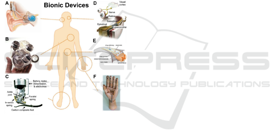
materials, blood, heart, etc (figure 1).
Figure 1: Bionic technologies for restorative medicine.
(A) Cochlear implant. (B) AbioCor self-
contained replacement heart. (C) Powered ankle-foot
prosthetic controlled by a neuromuscular model. (D)
Epiretinal, subretinal, and suprachoroidal implants.
(E) Electronic dura mater, ''e-dura,'' tailored for the
spinal cord. (F) A skin-inspired digital
mechanoreceptor, where the image shows a model
hand with DiTact sensors on the fingertips
connected with stretchable interconnects (Kong,
2016).
The development trend of materials is complex,
intelligent, dynamic, environmental, and bionic
materials have these aspects. The development and
achievements of bionic materials will affect all
aspects of society. It will bring about changes to the
human organs and biological systems and make the
materials and applications.
In short, the biomimetic medical materials
developed should have biocompatibility and
flexibility. They should contain cellular and
molecular induction and adhesion sites, sufficient
mechanical strength, and biodegradable and tissue
remodeling properties. To become a suitable
material for model biomedicine, the first
requirement is the ideal effect that is effective in the
body. The combination of biomaterials, technology,
software and equipment and interdisciplinary can
provide a systematic approach for medical
applications.
4 CONCLUSIONS
Four types of biomimetic medical materials with
different research have been discussed such as 1)
biomimetic materials of silk protein, 2) biomimetic
materials of nacre, 3) biomimetic materials of bone,
and 4) nano biomimetic materials. And these
materials can be applied to human medical research
at the medical level by transplanting some
recognizable characteristics in the body such as
cochlear implant, mechanical heart and so forth,
which is a boon for the disabled or the patient.
The development of this material involves many
cutting-edge disciplines and high-tech fields, such as
material science, medicine, cytology, engineering,
bionics, biotechnology, etc. If biomimetic medical
materials can get substantial and stable development,
they will have significant application prospects and
social effects. In the future, better biocompatibility,
biodegradability, good mechanical strength and
biological stability, and clinical applicability will
still require extensive and in-depth research.
REFERENCES
Almqvist, N., et al. (1999) Methods for fabricating and
characterizing a new generation of biomimetic
materials. Materials Science and Engineering: C. 7(1):
p. 37-43.
Altman, G.H., et al. (2002). Silk matrix for tissue
engineered anterior cruciate ligaments. Biomaterials.
23 (20): p. 4131-4141.
Applegate, M.B., et al. (2016). Photocrosslinking of silk
fibroin using riboflavin for ocular prostheses.
Advanced materials.28(12): p. 2417-2420.
Arcidiacono, S., et al.. (2002). Aqueous processing and
fiber spinning of recombinant spider silks.
Macromolecules. 35(4): p. 1262-1266.
Balasubramanian, V., et al. (2017). Biomimetic
Engineering Using Cancer Cell Membranes for
The Potential Application in Medical Biomimetic Materials
783

Designing Compartmentalized Nanoreactors with
Organelle-Like Functions. Advanced Materials.29(11):
p. 1605375.
Das, D. and I. Noh. (2018). Overviews of Biomimetic
Medical Materials, in Biomimetic Medical Materials:
From Nanotechnology to 3D Bioprinting, I. Noh,
Editor. Springer Singapore: Singapore. p. 3-24.
Gray, G.M., et al. (2016). Secondary Structure Adopted by
the Gly-Gly-X Repetitive Regions of Dragline Spider
Silk. International journal of molecular
sciences.17(12): p. 2023.
Heuer, A.H., et al. (1992). Innovative materials processing
strategies: a biomimetic approach. Science.255(5048):
p. 1098-1105.
Hinman, M.B., J.A. Jones, and R.V. Lewis. (2000).
Synthetic spider silk: a modular fiber. Trends in
biotechnology. 18(9): p. 374-379.
Kong, Y., et al. (2016). 3D Printed Bionic Nanodevices.
Nano Today. 11.
Lazaris, A., et al. (2002) Spider silk fibers spun from
soluble recombinant silk produced in mammalian cells.
science. 295(5554): p. 472-476.
Meinel, L., et al. (2005). Silk implants for the healing of
critical size bone defects. Bone.37(5): p. 688-698.
Melke, J., et al. (2016). Silk fibroin as biomaterial for
bone tissue engineering. Acta biomaterialia. 31: p. 1-
16.
Qian, C., et al. (2004). Super-hydrophobic characteristics
of butterfly wing surface. Journal of Bionic
Engineering.1(4): p. 249-255.
Qiaoling, H., et al. (2003) Studies on chitosan rods
prepared by in situ precipitation method. Chemical
Journal of Chinese Universities. 24(3): p. 528-531.
Shi, W., et al. (2017) Structurally and functionally
optimized silk-fibroin-gelatin scaffold using 3D
printing to repair cartilage injury in vitro and in vivo.
Advanced materials. 29(29): p. 1701089.
Tabata, Y.(2001). Recent progress in tissue engineering.
Drug discovery today. 6(9): p. 483-487.
Wang, Y. and X. Zhu.(2020).Nanofabrication within
unimolecular nanoreactors. Nanoscale. 12(24): p.
12698-12711.
Xu, Z., et al. (2015). Fabrication of a novel blended
membrane with chitosan and silk microfibers for
wound healing: characterization, in vitro and in vivo
studies. Journal of Materials Chemistry B. 3(17): p.
3634-3642.
Yan, Q., et al. (2013). Breathing Polymersomes: CO2-
Tuning Membrane Permeability for Size-Selective
Release, Separation, and Reaction. Angewandte
Chemie International Edition. 52(19): p. 5070-5073.
Yodmuang, S., et al. (2015). Silk microfiber-reinforced
silk hydrogel composites for functional cartilage tissue
repair. Acta biomaterialia.11: p. 27-36.
Yoo, S.C., et al. (2018). Biomimetic Artificial Nacre:
Boron Nitride Nanosheets/Gelatin Nanocomposites for
Biomedical Applications. Advanced Functional
Materials. 28 (51): p. 1805948.
ICBEB 2022 - The International Conference on Biomedical Engineering and Bioinformatics
784
