
Study of the Optical Properties of Biological Tissues Quantitative
Assessment Possibility using Spatial-Frequency
Domain Imaging
Alexander V. Kolpakov, Yuliya V. Ivanova, Svetlana A. Yushina and Anastasia S. Semenova
Department of Biomedical Technical Systems, Bauman Moscow State Technical University,
2-nd Baumanskaya str., 5/1, Moscow, Russia
Keywords: Optical Properties, Biological Tissues, Spatial-frequency Domain Imaging, Spectrophotometry, Optical
Imaging, Quantitative Assessment.
Abstract: The paper is devoted to the study of the possibility of using spatial-frequency domain imaging (SFDI)
technology in the tasks of visualizing neurovascular structures in the brain tissue during endosurgical
intervention, as well as the detection and objective quantitative assessment of lesions in the tissues of the
oral cavity. As a result of the initial stage of experimental studies in vivo, it was shown that SFDI allows
tracking the dynamics of optical parameters of tissues as a result of changes in blood filling and is
potentially applicable for detecting subsurface optical inhomogeneities in tissues.
1 INTRODUCTION
In recent decades, optical imaging has become
increasingly important in the field of medicine and
biology. The use of biological tissues optical
properties is widely used: from fundamental
research to obtain new knowledge about biological
processes to screening and numerical assessment of
the pathological processes stage.
The interest in optical methods is the result of
their unique property - non-invasiveness. The
sources of optical contrast are two main physical
phenomena - absorption and scattering of photons in
tissue.
One of the technologies for studying the optical
properties of tissue is the spatial frequency domain
imaging (SFDI). The main advantage of SFDI is the
ability to separate absorption and scattering effects
in the formation of optical contrast.
As a result of research in recent years, a number
of laboratories have shown the possibility of
visualizing the distribution and objective
quantitative assessment of the optical properties of
biological tissues using SFDI (Applegate, 2020;
Svaasand et al., 1999; Cuccia et al., 2019; Svaasand
et al., 1994; Cuccia et al., 2009; Hielscher et al.,
1975; Groenhuis et al., 1983; O'Sullivan et al., 2012;
Gioux et al., Carole et al., 2019).
At present, on the basis of the Department of
Biomedical Technical Systems of the Bauman
Moscow State Technical University, studies of SFDI
are being carried out, the purpose of which is to
determine the possibility of using SFDI in a number
of areas of medicine, in particular:
- in neurosurgery to solve the problem of
visualizing neurovascular structures in the
brain tissue during endosurgical intervention.
The hypothesis being tested is that due to the
peculiarity of SFDI, namely, due to the
possibility of separating with its help the
contribution of absorption and scattering to the
total attenuation of backscattered radiation,
there is the possibility of separate visualization
of blood vessels, as more absorbing structures,
and nerves, as more dissipative;
- in dentistry for visualization and quantitative
assessment at an early stage of inflammatory
processes, as well as erosive and ulcerative
lesions. The difference in the values of the
absorption and scattering coefficients of
intact and affected tissues of the oral cavity
can be hypothetically determined using SFDI
and used in the system for automatic
detection and control of the dynamics of
inflammatory and precancerous processes in
the tissues of the oral cavity at an early stage.
Kolpakov, A., Ivanova, Y., Yushina, S. and Semenova, A.
Study of the Optical Properties of Biological Tissues Quantitative Assessment Possibility using Spatial-Frequency Domain Imaging.
DOI: 10.5220/0010969300003123
In Proceedings of the 15th International Joint Conference on Biomedical Engineering Systems and Technologies (BIOSTEC 2022) - Volume 1: BIODEVICES, pages 317-321
ISBN: 978-989-758-552-4; ISSN: 2184-4305
Copyright
c
2022 by SCITEPRESS – Science and Technology Publications, Lda. All rights reserved
317
As a result of previously conducted research at
the Department of Biomedical Technical Systems:
- proved that the problem of detecting blood
vessels and cranial nerves during endoscopic
removal of tumors of the base of the skull can
be solved using the methods of automatic
detection of blood vessels and cranial nerves
based on methods of multispectral image
analysis and quantitative spectrophotometry
with local sensing in red and near infrared
(hereinafter - RNIR) wavelength ranges
(Safonova et al., 2019; Safonova et al., 2020;
Safonova et al., 2019; Safonova et al., 2019;
Safonova et al., 2019).
Under the conditions of the stand experiment the
possibility of detecting blood vessels located in the
tissues of the brain at a depth of up to 3 mm using
images of the red and near infrared range confirmed
(Kolpakov et al., 2021).
The possibility of visualization of the oral cavity
soft and hard tissues structure using radiation of the
near infrared wavelength range by diaphanoscopy has
been proven, as well as the possibility of detecting
lesions in the oral cavity tissues using visualization in
the near infrared range (Kolpakov et al., 2016).
SFDI is a new quantitative imaging technique, a
broadband diffuse optical technique that can separate
the effects of absorption and scattering and therefore
approximately determine the number of
chromophores in tissues. The method can be used to
measure the concentrations of tissue components
such as oxyhemoglobin, deoxyhemoglobin, lipids,
and water (Applegate et al., 2020).
Spatial frequency domain imaging consists of
projecting a two-dimensional (2-D) light pattern,
usually in the form of a harmonic periodic grating
(HPG), and analyzing the effect of multiple
scattering and absorption in tissue on the amplitude
of attenuated radiation, backscattered or past. In this
case, the amplitude of the attenuated radiation is
considered as a function of the spatial frequency of
the pattern.
2 MATERIALS AND METHODS
2.1 Stand for SFDI Research
To study the possibility of using SFDI, an
experimental stand is required. Currently, scientists
from Boston University (Applegate et al., 2020)
have created an openSFDI guide to create such a
stand from publicly available components
(hereinafter - the stand).
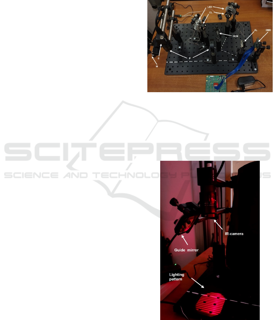
The stand contains three main modules: a
lighting module, a spatial modulation subsystem and
an image registration module (Figure 1).
Figure 1: The complete stand (off State). LED – light-
emitting diodes; CL - collimating lenses; DCM – dichroic
mirrors; AL - achromatic lens; P - linear polarizers; M –
guiding mirror; C - camera; DMD - digital micromirror
device.
An example of generating a lighting pattern is
shown in Figure 2. The object under study must be
placed in the illumination area.
Figure 2: Registration of the template (HPG) at the stand.
RMHM 2022 - Special Session on Remote Management and Health Monitoring
318
The programming of the controller with the
spatial modulator DMD was carried out in
accordance with the instructions for the openSFDI
system (Applegate et al., 2020).
To generate lighting patterns and register the
resulting images, we used software based on the
LabView platform, developed by a research group
from Boston University (Applegate et al., 2020).
The assembled stand allows image registration at
wavelengths of 660 and 850 nm at various spatial
radiation modulation frequencies.
The processing of experimental results in the
general case for SFDI technology consists of four
stages:
- demodulation over three images at the same
spatial frequency: with phases of 0, 120 and
240 degrees,
- calibration to separate the instrument
response function (IRF or MTF
system
) from the
response function of the test sample,
- determination of the diffuse reflection
coefficient of the recorded sample,
- determination of the optical characteristics of
the recorded sample as a result of solving the
inverse problem: from the known values of
the diffuse reflection coefficient R
d
at two (or
more) spatial frequencies, the values of the
optical parameters of the object are
determined, which satisfy the radiation
transfer equation.
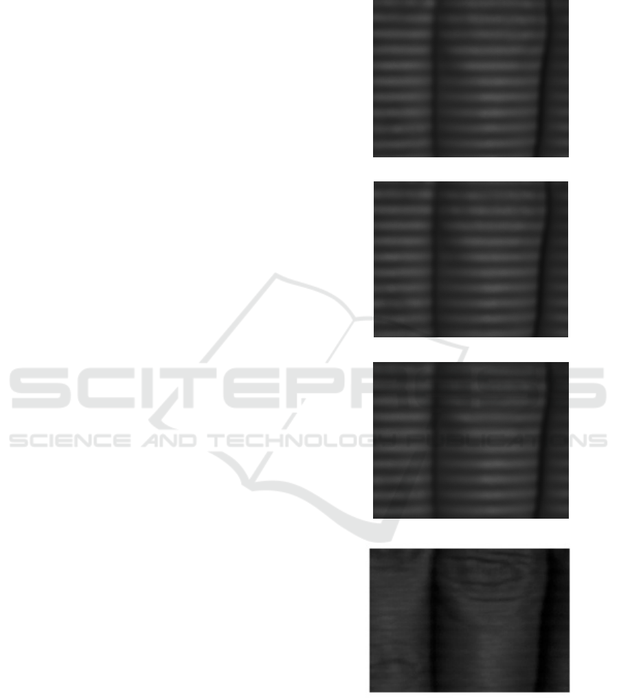
Figure 3 shows images with phases of 0, 120 and
240 degrees, obtained at the stand, as well as the
result of demodulation of these images. From three
images obtained in three phases, as a result of
demodulation, one image is obtained without stripes.
2.2 Initial Experimental Study
In order to initially test the possibility of measuring
the dynamics of biological tissues optical parameters
using SFDI, an experiment was carried out on the
assembled stand, during which the change in the
absorption and scattering coefficients on the surface
of the back of the volunteer's palm was monitored
during the change in the blood volume of the hand
using an occlusive cuff.
In the course of the experiment, the cuff was put
on a few centimeters above the bend of the subject's
elbow, and SFDI images of the back of the hand
were registered (Figure 4) at radiation wavelengths
of 660 and 850 nm in the non-occluded mode, in
venous oclussion mode (cuff pressure values 90 and
110 mm. Hg) and arterial occlusion mode (cuff
pressure value 220 mm Hg).
a)
b)
c)
d)
Figure 3: Images of the object taken at the stand.
a) -c ) Illumination with a spatial frequency of
0.5 mm
-1
, with phases of 0 °, 120 ° and 240 °;
d) result of demodulation.
Study of the Optical Properties of Biological Tissues Quantitative Assessment Possibility using Spatial-Frequency Domain Imaging
319
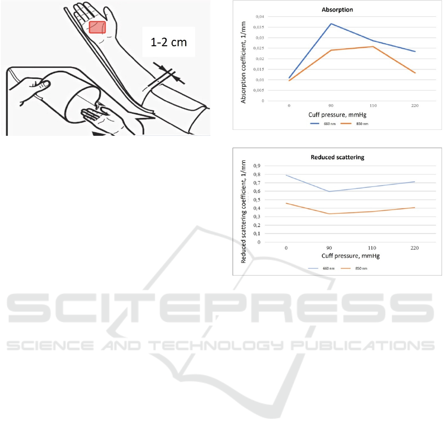
Figure 4: Scheme of SFDI images registration in an
experiment with an occlusal cuff: the area of registration is
marked with a red rectangle.
The registered images were used to determine
the values of the absorption and reduced scattering
coefficients averaged over the selected area.
3 RESULTS AND DISCUSSION
As a result of the initial work, a stand for
researching the SFDI technology was assembled.
The performance of the stand is confirmed by
obtaining a high-quality demodulated image.
The experimental dependences of the absorption
and reduced scattering coefficients on the pressure in
the occlusal cuff, obtained as a result of the
experiment, are shown in Figure 5.
The extrema of the values of the absorption and
reduced scattering coefficients were registered in the
mode of venous occlusion, corresponding to the
maximum blood filling of the investigated area. This
observation corresponds to the hypothetical
dynamics of the optical properties of the studied
tissues under the conditions of the experiment. Thus,
as a result of the analysis of the obtained
experimental dependences, it was found that the
SFDI technology makes it possible to adequately
monitor the dynamics of optical parameters of
tissues as a result of changes in blood filling and is
potentially applicable for the detection of subsurface
optical inhomogeneities in tissues, such as blood
vessels, inflammation foci.
To determine the accuracy of tissues optical
parameters SFDI measurements, it is required to
conduct a series of measurements on a larger sample
of subjects, as well as to develop SFDI metrological
support, including sets of measures, and methods for
verifying the results of SFDI measurements.
a)
b)
Figure 5: Parameters of absorption and scattering obtained
during the experiment on the stand: a) the value of the
absorption coefficient of the palm, depending on the
pressure in the cuff; b) the value of the reduced scaterring
coefficient of the palm depending on the pressure in the
cuff.
In the course of further work, it is planned:
- verification of the obtained experimental
dependences of the results of SFDI
measurements on the pressure in the occlusal
cuff on a larger number of subjects;
- verification of the results of SFDI
measurements using phase modulation
spectrophotometry;
- carrying out research at the stand in order to
determine the best values of the radiation and
registration parameters of the SFDI system
for the possibility of using it in the tasks of
visualizing neurovascular structures in the
brain tissue during endosurgical intervention,
detecting lesions in the tissues of the oral
cavity;
- development of metrological support for
SFDI systems;
- development, manufacture and clinical
testing of prototypes of SFDI systems for
visualization of neurovascular structures in
RMHM 2022 - Special Session on Remote Management and Health Monitoring
320

the brain tissue during endosurgical
intervention, detection and objective
numerical assessment of lesions in the tissues
of the oral cavity.
REFERENCES
Applegate M.B. (et al.). OpenSFDI: an open-source guide
for constructing a spatial frequency domain imaging
system // Journal of Biomedical Optics – 2020. – Vol.
25. No. 1.
OpenSFDI (Electronic resource). – Access mode:
http://opensfdi.org/ (date of the application:
23.04.2021).
Svaasand L., Spott T., Fishkin J. Reflectance
measurements of layered media with diffuse photon-
density waves: a potential tool for evaluating deep
burns and subcutaneous lesions // Physics in medicine
and biology – 1999. – Vol. 44. No. 3. P. 801 – 813.
Cuccia D., Gioux S., Mazhar A. Spatial frequency domain
imaging in 2019: principles, applications, and
perspectives // Journal of Biomedical Optics – 2019. –
Vol. 24. No. 7. P. 071613-1 – 071613-18.
Svaasand L., Haskell R., Tsay T. Boundary Conditions for
the Diffusion Equation in Radiative Transfer // Journal
of the Optical Society of America – 1994. – Vol. 11.
No. 10. P. 2727-2741.
Cuccia D., Bevilacqua F., Durkin A. Quantitation and
mapping of tissue optical properties using modulated
imaging // Journal of Biomedical Optics – 2009. – Vol.
4. No. 2. P. 024012-1 – 024012-13.
Hielscher A., Jacques S., Wag L. The influence of
boundary conditions on the accuracy of diffusion
theory in time-resolved reflectance spectroscopy of
biological tissues // Physics in medicine and biology –
1995. – Vol. 40. P. 1957 – 1975.
Groenhuis R., Ferwerda H., Ten Bosch J. Scattering and
absorption of turbid materials determined from
reflection measurements. 1: Theory // Journal of the
Optical Society of America – 1983. – Vol. 22. No. 16.
P. 2456-2462.
O'Sullivan T., Cerussi A., Tromberg B., Cuccia D. Diffuse
optical imaging using spatially and temporally
modulated light // Journal of Biomedical Optics –
2012. – Vol. 17. No. 7. P. 071311-1 – 071311-14.
Labview control software for the openSFDI platform
(Electronic resource). – Access mode:
https://github.com/mbapplegate/openSFDI-control
(date of the application: 24.04.2021)
Gioux S. (et al.) Spatial Frequency Domain Imaging in
2019: Principles, Applications and Perspectives //J
Biomed Opt. 2019; 24(7), 071613.
Carole K. H. (et al.). Optical sampling depth in the spatial
frequency domain // J. Biomed. Opt. 24(7), 071603
(2019), doi: 10.1117/1.JBO.24.7.071603.
Safonova L.P., Orlova V.G., Shkarubo A.N. Investigation
of Neurovascular Structures using Phase-Modulation
Spectrophotometry // Optics and Spectroscopy, 2019.
Vol.126. №6. P.820. doi: 10.1134/S0030400X19060
201
Orlova V. G., Safonova L. P., Soloveva P. M. In Vitro
Study of Optical Properties of the Central Nervous
System Components// EIConRus. St. Petersburg and
Moscow, 2020. P. 1567. DOI 10.1109/EIConRus49
466.2020.9039169
Safonova L. P., Orlova V. G., Lesnichaia A.D., Shkarubo
A.N., Chernov I.V. The Potential of the
Spectrophotometric Method for Detection and
Identification of Neurovascular Structures //
Biomedical Engineering 2019. Vol. 52 P. 402.
doi 10.1007/s10527-019-09856-6
Orlova V. G., Lesnichaia A.D., Safonova L. P. Algorithm
for Recognition of Vascular Structures in the Biotissue
Volume//Proceedings - 2019 Ural Symposium on
Biomedical Engineering, Radioelectronics and
Information Technology, Usbereit 2019. doi: 10.1109/
USBEREIT.2019.8736629
Al Khadzh K.A., Safonova L.P. The Research of the
Spectrophotometric Method for the Objective Control
of the Auditory Sense// Proceedings of the 2019 IEEE
Conference of Russian Young Researchers in
Electrical and Electronics Engineering, ElConRus
2019.DOI: 10.1109/EIConRus.2019.8657128
Yushina S.A., Kuznetsov N.N., Kolpakov A.V.
Investigation of the Possibility of Detecting Optical
Inhomogeneities in Brain Tissues from the Visible and
Near Infrared Images// Optics and Spectroscopy, 2021.
Vol.129.
№9.P.1204.doi: 10.21883/OS.2021.09.51352.1982-21
Kolpakov A.V., Judin I.N., Spiridonov I.N., Zorina O.A.
Early Detection of Inflammation Foci in the Soft
Tissues of the Periodontum using Infrared
Diaphanoscopy// Biomedical Engineering. 2016.
Vol.50.№2.p.88-91.
Study of the Optical Properties of Biological Tissues Quantitative Assessment Possibility using Spatial-Frequency Domain Imaging
321
