
Does Kinesio Taping Functional Correction Technique Affect
Walking Plantar Pressures?
Dalibor Kiseljak
1,2 a
, Filip Bolčević
1,2 b
and Vladimir Medved
1c
1
University of Zagreb, Faculty of Kinesiology, Horvaćanski zavoj 15, Zagreb, Croatia
2
University of Applied Health Sciences, Mlinarska cesta 38, Zagreb, Croatia
Keywords: Pedobarography, Kinesiotaping, Kinesiology, Biomechanics.
Abstract: The use of the Kinesio Taping (KT) method is widespread in sports, but current research on the ability of KT
to alter foot biomechanics is limited. The aim of this study was to examine the acute effects of KT on walking
plantar pressures. Kinesio Taping Functional Correction Technique was applied on the transverse arch of the
foot. Twenty-two young healthy male adults, aged between 19 and 33 voluntarily participated in this study.
Plantar pressures for three functional segments (forefoot, midfoot and hindfoot) were recorded during walking
(with and without KT) by pedobarographic measurement using FDM 1.5 pressure measuring device. The
results show that KT Functional Correction Technique has an effect in reducing walking plantar pressures. It
was shown that maximal plantar pressures at the forefoot and hindfoot significantly (P < 0.05) decreased by
1.51 N/cm
2
and 0.9 N/cm
2
respectively, when KT was applied to the foot during walking. KT has the potential
for primary and secondary prevention of pathological manifestations in the mentioned area, but also in the
holistic postural context. Further research is needed to investigate clinical significance of KT.
1 INTRODUCTION
There are various kinematic and kinetic tasks of the
ankles and feet, from contact with the ground and
adaptation to different surfaces, through deceleration,
shock absorbing, to propulsion necessary for bipedal
locomotion. High forces are transmitted through the
most distal segments of the lower extremities. The
arches of the foot frequently participate in the
absorption and distribution of forces (Houglum and
Bertoti, 2012). In a normal foot the segments of the
lateral arch at full contact with the load of the foot
(closed kinetic chain) are always in contact with the
ground, while in a normally aligned foot the medial
arch is rather high. The third arch of the foot is
transverse, extending from medial to lateral through
the cuneiform bones to the cuboid bone. Together
with the longitudinal arches, it enables efficient load
absorption from the superior direction (body weight),
but also from the inferior one (ground reaction forces).
Along with the joint congruence of the middle part of
the foot, the transverse arch is maintained by the
a
https://orcid.org/0000-0003-2659-5949
b
https://orcid.org/0000-0002-6588-2468
c
https://orcid.org/0000-0002-8298-5602
intrinsic muscles. Biomechanical integrity of the foot
is achieved through the alignment of bone and joint
segments, stabilization of plantar ligaments, support
of plantar fascia and by intrinsic and extrinsic
muscles. These structures act together to absorb the
ground reaction forces (Oatis, 2009).
Foot is functionally divided into three parts:
hindfoot, midfoot and forefoot. Hindfoot includes
talus and the first segment of contact with the ground
at the initial stance phase in the gait cycle - calcaneus.
Main midfoot function in the relationship between
stability and mobility is transfer of closed kinetic
chain movements and related forces through
tarsometatarsal joints to the forefoot, a segment
crucial for terminal stance and pre-swing which is
generally extremely adaptable to ground contact
through various forms of closed kinetic chain
(Houglum and Bertoti, 2012). Weight distribution in
the foot can affect the bearing line of the ankle, knee,
and hip (Guner and Alsancak, 2020).
Pedobarography is a modern technology enabling
the assessment of the locomotor system based on the
Kiseljak, D., Bol
ˇ
cevi
´
c, F. and Medved, V.
Does Kinesio Taping Functional Correction Technique Affect Walking Plantar Pressures?.
DOI: 10.5220/0011379300003321
In Proceedings of the 10th International Conference on Sport Sciences Research and Technology Support (icSPORTS 2022), pages 71-77
ISBN: 978-989-758-610-1; ISSN: 2184-3201
Copyright
c
2022 by SCITEPRESS – Science and Technology Publications, Lda. All rights reserved
71

plantar pressure distribution. Also, this technique is
useful in the rehabilitation of various types of
dysfunctions of body movement (Lorkowski and
Gawronska, 2021). Therefore, in assessment of data
about feet structure and function kinetic and
kinematic pressure measurement devices (PMDs) are
used (Gruić et al., 2015). PMDs measure plantar
pressure distribution beneath the foot to quantify
pressure distribution, pressure magnitude and
progression of the centre-of-pressure (CoP) as the
participant walks barefoot over a force plate
embedded in a walkway (Jameson et al., 2008). In
contrast to motion capturing systems,
pedobarography can usually be completed in shorter
time and requires less specialized expertise to be
performed. Anyway, pedobarography is ideally used
with motion capture type analysis to provide
complementary information on foot dynamics.
However, when foot posture is the primary area of
interest, pedobarography may provide sufficient
information to answer clinical questions (Mudge et al.,
2020). In addition to clinical examination of the
patient, in this manner we get very useful information
about the state of foot and the type of load in certain
phases of walking. Data collection must be
standardized so that they can be analysed and follow
the results for each patient, as well as compare them
with certain standards. In diagnosis pedobarography
is used in case of walk disorders after surgery of the
hip and knee, stroke, or other neurological problems.
It is important to highlight the clinical application of
pedobarography in diabetology, sports medicine and
treatment of foot deformities and rehabilitation
(Skopljak et al., 2014). It is known that the plantar
surface of the foot is the most common place for
occurrence of foot ulcerations. During ordinary or
sport activities the foot is exposed to high static and
dynamic loading forces, which can lead to
disharmony of muscle strength and load which lead
to appearance of overuse injuries. Accordingly, there
are many strategies to reduce the maximal pressure
during walking through relief of total contact with
insoles or specifically prescribed foot and ankle
exercises (Choi et al., 2014). Kinesio Taping (KT) has
gained popularity in the treatment of musculoskeletal
pathologies (Griebert et al., 2016). Regarding the
prevention and rehabilitation of ankle and foot
dysfunction, this method has been widely used in
sports in recent years (Aguilar et al., 2016).
KT is an active, not a passive method; according
to Aktas and Baltaci (2011), Kinesio tape does not act
as an immobilizer, but through the improvement of
proprioception (de Oliveira et al., 2019) provides
active support to the musculoskeletal system.
Therefore, KT is significantly different from Athlet
Taping (AT) or orthoses. According to Yen et al.
(2018), KT is significantly superior to AT in order to
dynamically reduce foot inversion (or to increase foot
eversion) during early stance phase. Self-adhesive,
longitudinally elastic, cotton Kinesio tape is more
porous and waterproof than standard bandage tapes,
which allows it to be worn for 3 to 5 days after
application (Kase et al., 2013). There are no medical
substances on the Kinesio tape. The principle of its
action is based on mechanical properties, primarily on
elasticity (Kahanov, 2007) which is approximately
50% of the initial length, which is equivalent to the
elasticity of the skin (Kase et al., 2013). The tendency
towards active, mobilization, movement is contained
in the name of the method, which is derived from the
term "kinesiology", since the use of Kinesio tape
seeks to enable normal movement of the body and
body segments (Kase et al., 2013).
Optimization of muscle function and correction of
malaligned joints are the effects of KT (Kase et al.,
2013) which can be very useful for the purpose of
improving posture and movement even among
asymptomatic individuals. The neuromuscular basis
of KT application implies the ability to improve
proprioception (Kurt et al., 2016), a mechanism that
Aarseth et al. (2015) describe as an increase in muscle
pressure due to increased mechanoreceptor
stimulation in the skin, which affects joint mechanics.
According to Kase et al. (2013), the KT method can
be applied at any stage of the rehabilitation process,
but also in primary and/or secondary prevention
where, as interpreted by Lemos et al. (2015), KT can
be an input for solving a closed circle of pathological
and compensatory patterns of posture and movement.
Because the foot has a key function in absorbing and
transmitting forces throughout the body, structural
and functional postural deficiencies in the ankle and
foot area can create repercussions on other segments
of the kinetic chain, even those very distant. Thus, for
example, collapsed arch of the foot could cause neck
pain as a symptomatic manifestation of compensatory
changes of the cervical spine and vertebral column
and trunk, following the cause-and-effect relationship
(Page et al., 2010) of human posture. The results of
trial made by Aguilar et al. (2016) suggest that there
is some benefit in applying KT before intense
physical activity to change foot posture. According to
Griebert et al. (2016) KT can correct biomechanical
factors that may be associated with musculoskeletal
pathology. Yen et al. (2018) state that KT can be a
useful tool for correcting aberrant motion without
restricting natural movement in sports. On the other
hand, some researchers (Pérez-Soriano et al., 2014;
icSPORTS 2022 - 10th International Conference on Sport Sciences Research and Technology Support
72

Cornwall et al., 2019; Guner and Alsancak, 2020)
conclude that despite its widespread use, current
research potential of KT to change foot posture and
movement is limited.
In the available literature, we did not find any
research on the effect of KT on walking plantar
pressures after application of the KT Functional
Correction Technique as a support on the transverse
arch of the foot. In their prospective cohort study
published in 2019, where the KT muscle facilitation
technique was applied, Cornwall and colleagues
emphasized that additional studies need to be
conducted, with respect to other KT application
methods, to change plantar pressures.
Therefore, the aim of this study was to examine
whether KT Functional Correction Technique on the
transverse arch of the foot can improve dynamic
posture through the modification of walking plantar
pressures. The hypothesis was that walking maximal
plantar pressures would be significantly reduced after
the intervention for all three parts of the foot (forefoot,
midfoot and hindfoot), which we consider a positive
effect of KT.
2 METHODS
2.1 Participants
The convenience sample comprised of 22 healthy
male university students, with a mean age of 23.2 ±
3.6 years (ranging from 19 to 33 years), body height
178.5 ± 5.4 cm (ranging from 169.5 to 188.5 cm), and
a mean body weight 80.6 ± 11.7 kg (ranging from
60.2 to 101.6 kg). Inclusion criteria were no pain and
no history of lower limb injuries in the last 12 months.
All subjects participated in the study voluntarily.
Before enrollment, everyone was given informed
consent, which they had to sign as a prerequisite for
participating in the research. The study was approved
by the ethics committee of the Faculty of
Kinesiology, University of Zagreb, and was carried
out in accordance with the Declaration of Helsinki.
2.2 Procedure
The research was performed in the Biomechanics
Laboratory of Faculty of Kinesiology, University of
Zagreb. Measurement protocol started from initial
standing position with participants being barefoot.
Each participant walked over the trackway 9.5 m long
to the end of the trackway, turned around and went
back for 6 times. During the gait, subject should be
instructed to develop and reach velocity normal for
aiming himself towards ordinary activity when there
are no disturbing aspects.
The initial measurement was performed without
KT, and the final with KT (within-group design).
After the initial measurements, KT was applied
according to the protocol of Kase et al. (2013), and
after 60 min the acute effects of the intervention were
examined. The 60-min interval between intervention
and final testing (as in Donec et al., 2012 and Voglar
and Sarabon, 2014) increased the potential that the
material would be successfully applied, as discussed
by Aktas and Baltaci (2011) and Ruggiero et al.
(2016).
2.2.1 Instrument
The instrument used in this study was Zebris FDM
1.5 (Germany) gait analysis system which was
centrally positioned on a 158 cm long and 60.5 cm
wide trackway platform. Measurements on platform
are supported by 11264 capacitive sensors with
density of 1.4 sensors/cm² with measuring range 1-
120 N and accuracy ± 5%. Sensor area is 149 x 54.2
cm (L x W) with sampling rate 100 Hz.
2.2.2 Pedobarographic Measurement
Measurement and evaluation was done on the
personal computer using the intuitive Zebris FDM
Software Suite. It synchronously evaluates the
measuring data of the ground reaction forces and the
video camera. After defining the left and right ground
contacts, an analysis of the measuring cycles is
automatically performed in the subsequent report that
displays the measured results. Reports offer 63
quantitative variables and graphics within
participants like pressure plots, gait parameters
(geometry, phases, timing), CoP analysis, force and
pressure parameters, and curves, and three foot zone
analysis.
Maximal plantar pressures within each foot region
(forefoot, midfoot, hindfoot) were recorded
bilaterally for further data analysis. Walking speed
(velocity) was also measured.
2.2.3 Kinesio Taping Application
KT interventions were conducted by one researcher,
the author of this paper (master physiotherapist with
14 years of experience, also a certified Kinesio
Taping practitioner (CKTP) and instructor (CKTI)
with 11 years of experience with the KT method),
always through the same, standardized procedure
recommended by Kase et al. (2013). The application
of Kinesio tape was preceded by cleaning the skin
Does Kinesio Taping Functional Correction Technique Affect Walking Plantar Pressures?
73
with alcohol and placing the subjects in the prone,
with ankle in neutral position. The same material
Kinesio Tex Gold FingerPrint Tape, Kinesio Holding
Company, Albuquerque NM, 5 cm wide, black, was
used for all participants in the study. The KT
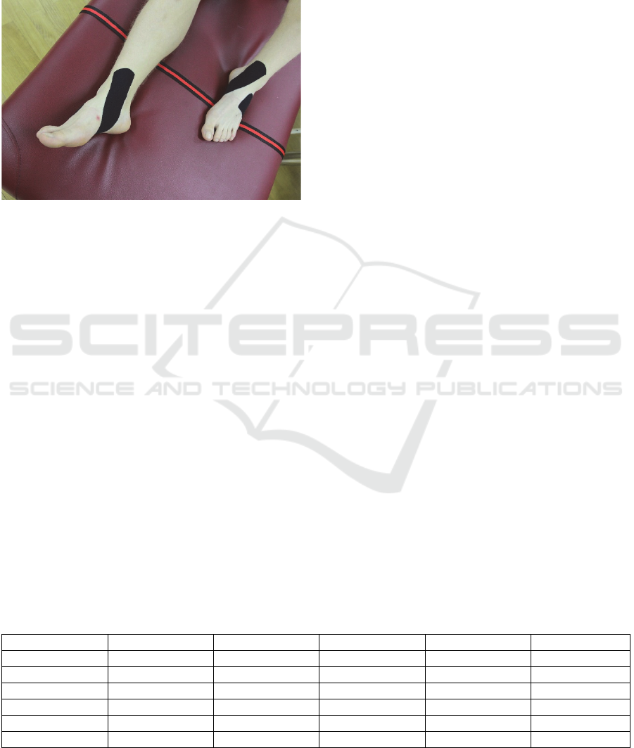
application is shown in Figure 1.
Figure 1: Kinesio Taping Functional Correction Technique
applied to the transverse arch of the foot.
Functional Correction Technique was chosen,
starting on lateral foot with no tension, then 75+ %
tension was applied across the arch (symbol + means
that tension is applied through movement). Finally,
end with no tension was applied on the medial aspect
of the foot. The adhesive on the tape was heat
activated by gently rubbing from the ends towards the
middle of the tape. KT Functional Correction
Technique is characterized by a “Spring-Assist or
Limit” mechanism that provides sensory stimulation
to either assist transverse arch and limit a motion
(arch collapse) by increasing stimulation to joint
receptors and mechanoreceptors.
2.3 Statistical Analysis
Data were analysed using the software package
STATISTICA v.13.5 (StatSoft, Inc., Tulsa, OK,
USA). Descriptive parameters were calculated, while
the main analysis comparing the condition without
KT (-) and the condition with KT (+) was performed
using the nonparametric Wilcoxon Matched Pairs
Test, with the timepoint as a dependent factor. The
level of statistical significance was set at P ≤ 0.05.
3 RESULTS
The main descriptive results of the assessment of
walking plantar pressure according to foot segments
and timepoints are presented in Table 1. Among the
results we can notice a decrease in walking maximal
plantar pressure after the intervention in all parts of
the foot.
The results of the Wilcoxon Matched Pairs Test
for the plantar pressure measured in this trial are
presented in Table 2. Statistically significant
differences (P < 0.05) were found for forefoot and
hindfoot walking maximal plantar pressures.
In order to check whether the walking speeds were
balanced according to the observed conditions (with
and without the KT), we applied Student’s t-test for
independent samples. The average walking speed of
the subjects in the condition with KT it was 4.5 ± 0.52
km/h, while in the condition without KT it was 4.57
± 0.42 km/h. No statistically significant differences in
walking speed (P = 0.592) were found between the
two conditions.
4 DISCUSSION
The aim of this study was to examine the acute effects
of KT on walking plantar pressures. The research
hypothesis was tested by checking the significance of
the dependent factor. It was shown that maximal
plantar pressures at the forefoot and hindfoot
significantly (P < 0.05) decreased by 1.51 N/cm
2
and
0.9 N/cm
2
respectively, when KT Functional
Correction Technique was applied to the foot during
walking.
Table 1: Descriptive forefoot, midfoot and hindfoot walking maximal plantar pressure (N/cm
2
) parameters, without (-) and
with (+) KT.
Variable N M SD Min Max
forefoot - 44 46.90 9.34 21.50 64.30
forefoot + 44 45.39 9.10 22.80 66.00
midfoot - 44 14.45 6.99 5.50 37.30
midfoot + 44 14.41 6.96 6.10 32.50
hindfoot - 44 35.77 7.41 22.10 49.30
hindfoot + 44 34.87 7.22 22.20 48.30
icSPORTS 2022 - 10th International Conference on Sport Sciences Research and Technology Support
74

Table 2: Wilcoxon Matched Pairs Test for forefoot, midfoot and hindfoot walking maximal plantar pressure, for condition
without (-) and with (+) KT.
Pair of Variables N T Z P
- and + (forefoot) 44 234.50 2.87 0.003979
- and + (midfoot) 44 483.00 0.14 0.888627
- and + (hindfoot) 44 326.50 1.96 0.049250
The post-intervention manifestation of the
“Spring-Assist or Limit” mechanism may be
associated with the activation of the central generator
of segmental posture and movement pattern via
proprioceptors, through a stimulus that acts on the
principle of pre-stressing (Kase et al., 2013).
As a result of its proposed therapeutic effect (Kase
et al., 2013), KT can be useful to people with
excessive foot pronation to improve function. Several
studies (Luque-Suarez et al., 2014; Pérez-Soriano et
al., 2014; Aguilar et al., 2016; Griebert et al. 2016;
Yen et al., 2018; Cornwall et al., 2019; Guner and
Alsancak, 2020) investigated the effects of KT in the
ankle and foot dysfunctions prevention and/or
rehabilitation. Among the mentioned studies, our
results are most consistent with the findings of
Griebert et al. (2016) who found that KT significantly
decreases plantar pressures. They analysed subjects
with Medial Tibial Stress Syndrome, an overuse
injury typical for physically active. We also find an
association with our results in a study by Yen et al.
(2018) which included a kinematic assessment of the
gait of individuals with chronic ankle instability.
Researchers found less foot inversion when walking
with KT compared to walking without KT during the
loading response phase.
Our results contrast with those of Aguilar et al.
(2016) who concluded that the application of KT on
the arch of the foot in healthy individuals has no
short-term effect on the change of walking plantar
pressures. Contrary to us, Aguilar et al. (2016) used
the KT Mechanical Correction Technique with 75%
tape stretch. The same KT technique and principles of
application were used by Guner and Alsancak (2020),
although they assessed the static load and concluded
that immediately after the application KT does not
alter weight bearing on the foot. Just the load-bearing
line of the ankle joint changed. Researchers conclude
that KT may be of some benefit in short-term
correction of foot pronation, although their practical
suggestion is to combine KT with orthotic footwear.
Luque-Suarez et al. (2014) applied tape with 100%
stretch, but their static kinematic assessment did not
show any postural improvement 24 hours after KT
application, compared to sham KT (applied without
tension). Contrary to our findings, according to
research by Cornwall et al. (2019) and Pérez-Soriano
et al. (2014), application of KT did not result in a
change in plantar pressure in healthy individuals.
Cornwall et al. (2019) applied the KT Muscle
Facilitation Technique for posterior tibialis muscle.
However, they made a big mistake by repeated
measurements only 5-10 min post-interventional,
since it takes a minimum of 30 min (Kase et al., 2013)
for KT to be effective with respect to adhesive
activation and adaptation. Regardless of this
oversight, the fact is that the arch of the foot is
maintained by the shape of the bones and their
interrelationships, furthermore by non-contractile
soft tissues (e.g. plantar ligaments and fascia) and
contractile soft tissues (i.e. muscles), where non-
contractile tissues make a greater contribution to arch
maintenance than contractile (Oatis, 2009).
Therefore, the KT Functional Correction Technique,
in our opinion, was a better choice for reducing
walking plantar pressures than the KT Muscle
Facilitation Technique. Pérez-Soriano et al. (2014)
applied KT on peroneus and triceps surae muscles,
examining changes in walking plantar pressures
(unlike Cornwall et al. (2019) who examined static
plantar pressures). Pérez-Soriano et al. (2014) discuss
that the walking pattern in terms of plantar pressure
distribution is not affected by KT Muscle Facilitation
Technique. Given the great forces required to
maintain the arch of the foot, in which non-contractile
structures predominate (Oatis, 2009), the KT Muscle
Facilitation Technique indeed seems too weak to
change plantar pressures. This goal requires a
technique that will not rely on the recoil mechanism
(in which the elasticity of the tape causes tissue
decompression (Tu et al., 2016)), but on the spring
mechanism in the function of limiting unwanted
movement or unwanted changes in segmental foot
posture - and that is characteristic of KT Functional
Correction Technique.
Our findings could be clinically relevant because
KT is a common method that is widely used by
various practitioners (e.g. physiotherapists, medical
doctors and athletic trainers), to prevent and/or
rehabilitate neuro-musculoskeletal disorders.
Following the premise of Luque-Suarez et al. (2014)
on KT as a simple alternative to traditional taping in
people with overpronated feet, we also see the
practical implications in the perspective of
Does Kinesio Taping Functional Correction Technique Affect Walking Plantar Pressures?
75

alternatives to orthopaedic insoles that provide
passive support, while KT Functional Correction
Technique with its “Spring-Assist or Limit”
mechanism is active. To the best of our knowledge,
this is the first study on the use of KT Functional
Correction Technique in plantar pressures analysis.
One strength of this study is that it was conducted
by an experienced physiotherapist and Certified
Kinesio Taping Instructor. Guided by the idea that KT
research should focus on the impact of the KT method,
and not on testing the effect of Kinesio tape placed on
the subject’s skin, with an emphasis on who, how and
for what purpose applies the tape, we fully agree with
Stockheimer et al. (2016) commentary “Research
requires deep knowledge of the modality to be tested”,
with universal repercussions, emphasizing the need
for adequate theoretical and practical education (i.e.
with certificates, licenses) of researchers who, in this
case, apply Kinesio tape, or rather apply the KT
method. We used a within-group design since
differences between groups in subject characteristics
could potentially negatively influence the results.
Nonetheless, the absence of a sham-tape group can be
considered a lack of the research as the role of placebo
effect regarding the use of KT is not investigated.
Furthermore, limitations of the current study are that
only acute effects of KT were assessed, and
longitudinal arches were not supported, considering
that the medial longitudinal arch is crucial (Oatis,
2009) for a normally aligned foot. A key limitation of
pedobarography is its inability to detect a patient’s
habit of avoiding pressure in the area of pain that
leads to an antalgic gait. An altered gait pattern can
affect pressure scores and provide contradictory
information on areas of pain (Choi et al., 2014).
Therefore, in future research, kinematic
assessment could be included as well as to evaluate
the impact of KT on foot biomechanics in a clinical
sample. Jumps or some sport-specific movements that
are subject to perturbations (Briem et al., 2011) and
require good proprioception as a risk zone for ankle
and foot injuries, could also be studied. Regarding the
clinical significance of the research, we agree with
Yen et al. (2018) that due to the small magnitude of
acute positive change, the clinical significance of our
results in terms of reducing the risk of injury is
unclear and should be investigated in the future,
through randomized clinical trials including a larger
sample size.
5 CONCLUSION
This study showed that Kinesio Taping method has a
positive effect on walking plantar pressures of healthy
individuals. Application of the Functional Correction
Technique significantly reduced walking maximal
plantar pressures in forefoot and hindfoot.
REFERENCES
Aarseth, L. M., Suprak, D. N., Chalmers, G. R., Lyon, L. &
Dahlquist, D. T. (2015). Kinesio tape and shoulder-joint
position sense. Journal of Athletic Training, 50(8), 785-
791.
Aguilar, M. B., Abián-Vicén, J., Halstead, J., & Gijon-
Nogueron, G. (2016). Effectiveness of neuromuscular
taping on pronated foot posture and walking plantar
pressures in amateur runners. Journal of science and
medicine in sport, 19(4), 348-353.
Aktas, G. & Baltaci, G. (2011). Does kinesiotaping increase
knee muscles strength and functional performance?.
Isokinetics and Exercise Science, 19(3), 149-155.
Briem, K., Eythörsdöttir, H., Magnúsdóttir, R. G.,
Pálmarsson, R., Rúnarsdöttir, T., & Sveinsson, T.
(2011). Effects of kinesio tape compared with
nonelastic sports tape and the untaped ankle during a
sudden inversion perturbation in male athletes. Journal
of Orthopaedic & Sports Physical Therapy, 41(5), 328-
335.
Choi, Y. R., Lee, H. S., Kim, D. E., Lee, D. H., Kim, J. M.,
& Ahn, J. Y. (2014). The diagnostic value of
pedobarography. Orthopedics, 37(12), e1063-e1067.
Cornwall, M. W., Jain, T. K., Holmgren, S., Dorri, A., &
Young, C. (2019). The effect of Kinesio Tape® on
static foot posture, plantar pressure, and rearfoot motion
in individuals with pronated feet. International Journal
of Sports Physical Therapy, 14(3), 368-375.
de Oliveira, F. C. L., de Fontenay, B. P., Bouyer, L. J. &
Roy, J. S. (2019). Immediate effects of kinesiotaping on
acromiohumeral distance and shoulder proprioception
in individuals with symptomatic rotator cuff
tendinopathy. Clinical Biomechanics, 61, 16-21.
Donec, V., Varžaitytė, L., & Kriščiūnas, A. (2012). The
effect of Kinesio Taping on maximal grip force and key
pinch force. Polish Annals of Medicine, 19(2), 98-105.
Griebert, M. C., Needle, A. R., McConnell, J., & Kaminski,
T. W. (2016). Lower-leg Kinesio tape reduces rate of
loading in participants with medial tibial stress
syndrome. Physical therapy in sport, 18, 62-67.
Gruić, I., Cebović, K., Radaš, J., Bolčević, F., & Medved,
V. (2015). Pedobarographic Features of Gait Measured
by FDM1. 5 PMD. In Proceedings of the 3rd
International Congress on Sport Sciences Research and
Technology Support (icSPORTS, 2015), 66-71.
SCITEPRESS.
Guner, S., & Alsancak, S. (2020). Kinesiotaping
Techniques to Alter Static Load in Patients With Foot
icSPORTS 2022 - 10th International Conference on Sport Sciences Research and Technology Support
76

Pronation. Journal of Chiropractic Medicine, 19(3),
175-180.
Houglum, P. A., & Bertoti, D. B. (2012). Brunnstrom's
clinical kinesiology. FA Davis Company. Philadelphia
(PA), 6
th
edition.
Jameson, E. G., Davids, J. R., Anderson, J. P., Davis, R. B.,
Blackhurst, D. W., & Christopher, L. M. (2008).
Dynamic pedobarography for children: use of the center
of pressure progression. Journal of Pediatric
Orthopaedics, 28(2), 254-258.
Kahanov, L. (2007). Kinesio Taping®, part 1: an overview
of its use in athletes. International Journal of Athletic
Therapy and Training, 12(3), 17-18.
Kase, K., Wallis, J., & Kase, T. (2013). Clinical
Therapeutic Applications of the Kinesio Taping
Method. 3rd ed. Albuquerque, NM: Kinesio Taping
Association International.
Kurt, E. E., Büyükturan, Ö., Erdem, H. R., Tuncay, F. &
Sezgin, H. (2016). Short-term effects of kinesio tape on
joint position sense, isokinetic measurements, and
clinical parameters in patellofemoral pain syndrome.
Journal of Physical Therapy Science, 28(7), 2034-
2040.
Lemos, T. V., Pereira, K. C., Protássio, C. C., Lucas, L. B.
& Matheus, J. P. C. (2015). The effect of Kinesio
Taping on handgrip strength. Journal of Physical
Therapy Science, 27(3), 567-570.
Lorkowski, J., & Gawronska, K. (2021). Pedobarography
in Physiotherapy: A Narrative Review on Current
Knowledge. In Integrative Clinical Research. Advances
in Experimental Medicine and Biology, 1375, 13-22.
Springer.
Luque-Suarez, A., Gijon-Nogueron, G., Baron-Lopez, F. J.,
Labajos-Manzanares, M. T., Hush, J., & Hancock, M.
J. (2014). Effects of kinesiotaping on foot posture in
participants with pronated foot: a quasi-randomised,
double-blind study. Physiotherapy, 100(1), 36-40.
Mudge, A. J., Sangeux, M., Wojciechowski, E. A., Louey,
M. G., McKay, M. J., Baldwin, J. N., Dwan, L. N., Axt,
M. W., & Burns, J. (2020). Can pedobarography predict
the occurrence of heel rocker in children with lower
limb spasticity?. Clinical Biomechanics, 71, 208-213.
Oatis, C. A. (2009). Kinesiology: the mechanics and
pathomechanics of human movement. Lippincott
Williams and Wilkins. Baltimore (MD), 2
nd
edition.
Page, P., Frank, C. C., & Lardner, R. (2010). Assessment
and Treatment of Muscle Imbalance: The Janda
Approach. Human Kinetics. Champaign (IL).
Pérez-Soriano, P., Lucas-Cuevas, A. G., Aparicio-Aparicio,
I., & Llana-Belloch, S. (2014). Effects of Kinesiotape®
taping on plantar pressure and impact acceleration
during walking. Science & sports, 29(5), 282-287.
Ruggiero, S. A., Frost, L. R., Vallis, L. A., & Brown, S. H.
(2016). Effect of short-term application of kinesio tape
on the flexion-relaxation phenomenon, trunk postural
control and trunk repositioning in healthy females.
Journal of Sports Sciences, 34(9), 862-870.
Skopljak, A., Muftic, M., Sukalo, A., Masic, I., & Zunic, L.
(2014). Pedobarography in diagnosis and clinical
application. Acta Informatica Medica, 22(6), 374-378.
Stockheimer, K. R., Baltaci, G., Forrester, G. G., Frassine,
S., & Wolkenberg, A. (2016). Research requires deep
knowledge of the modality to be tested. Journal of
Physiotherapy, 62(2), 118.
Tu, S. J., Woledge, R. C., & Morrissey, D. (2016). Does
‘Kinesio tape’ alter thoracolumbar fascia movement
during lumbar flexion? An observational laboratory
study. Journal of Bodywork and Movement Therapies,
20(4), 898-905.
Voglar, M., & Sarabon, N. (2014). Kinesio taping in young
healthy subjects does not affect postural reflex reactions
and anticipatory postural adjustments of the trunk: a
pilot study. Journal of Sports Science & Medicine,
13(3), 673-679.
Yen, S. C., Folmar, E., Friend, K. A., Wang, Y. C., & Chui,
K. K. (2018). Effects of kinesiotaping and athletic
taping on ankle kinematics during walking in
individuals with chronic ankle instability: A pilot study.
Gait & posture, 66, 118-123.
Does Kinesio Taping Functional Correction Technique Affect Walking Plantar Pressures?
77
